
What if a heart valve replacement could repair, adapt, and grow like living tissue?
Heart valve replacement has transformed the treatment of severe valve disease. Yet every existing replacement involves a compromise.
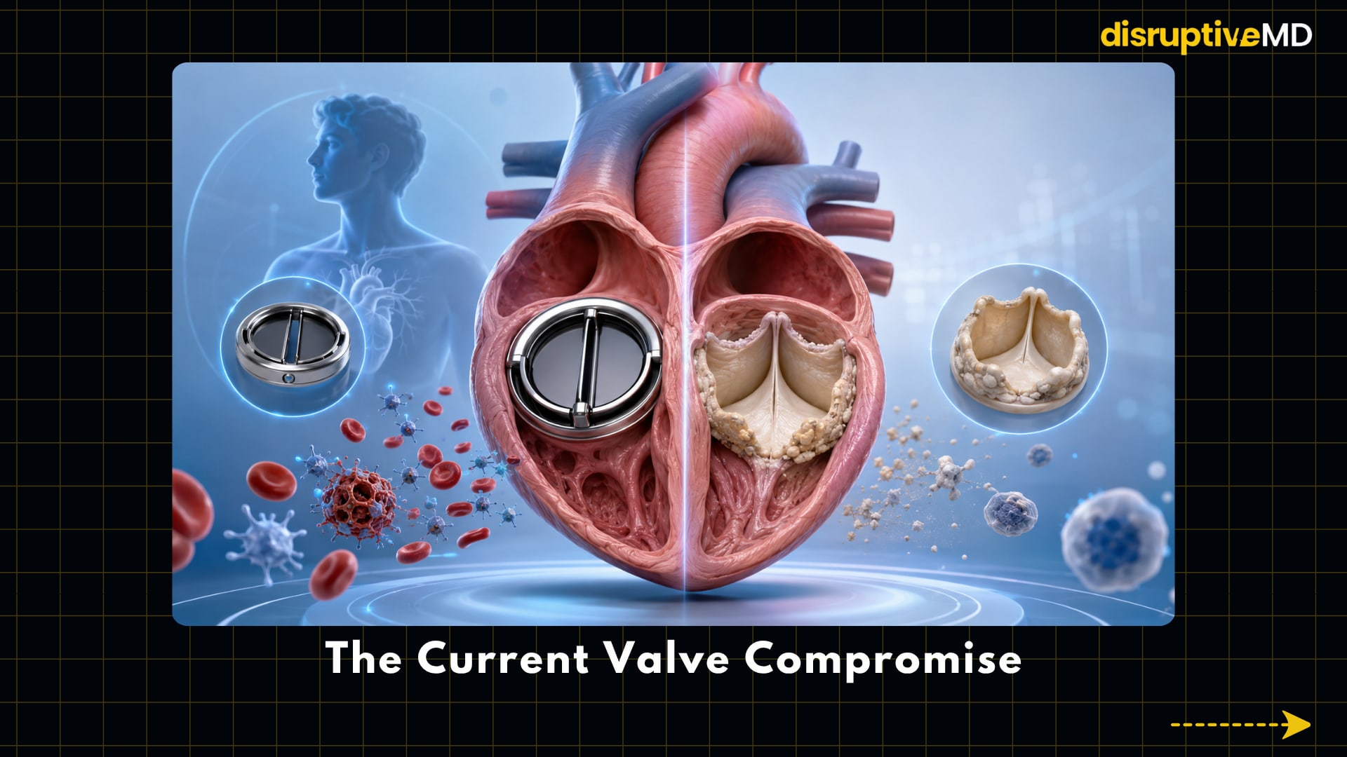
Mechanical valves offer exceptional durability, but most patients require lifelong anticoagulation to prevent dangerous blood clots. Biological valves—usually made from animal or donated human tissue—often avoid permanent anticoagulation, but they can stiffen, calcify and deteriorate over time. Their durability may be particularly limited in younger patients.
Bioengineered heart valves aim to move beyond this trade-off.
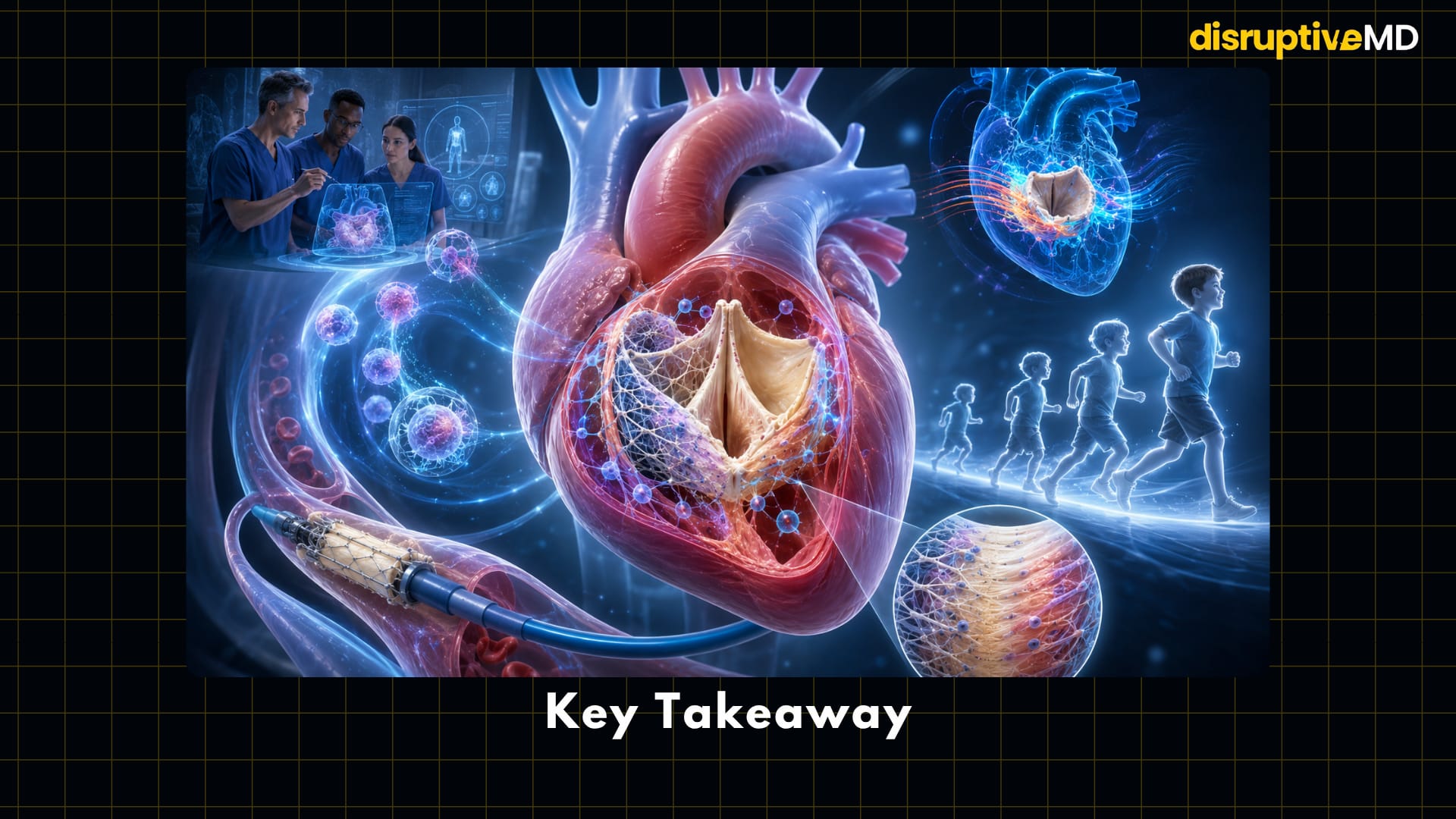
Instead of placing a permanently artificial device inside the heart, researchers are working toward valves that interact with the patient’s biology. These valves could recruit cells, remodel their own structure, resist calcification and potentially grow as a child grows.
The long-term vision is not simply a better prosthesis. It is a valve that gradually becomes part of the body.
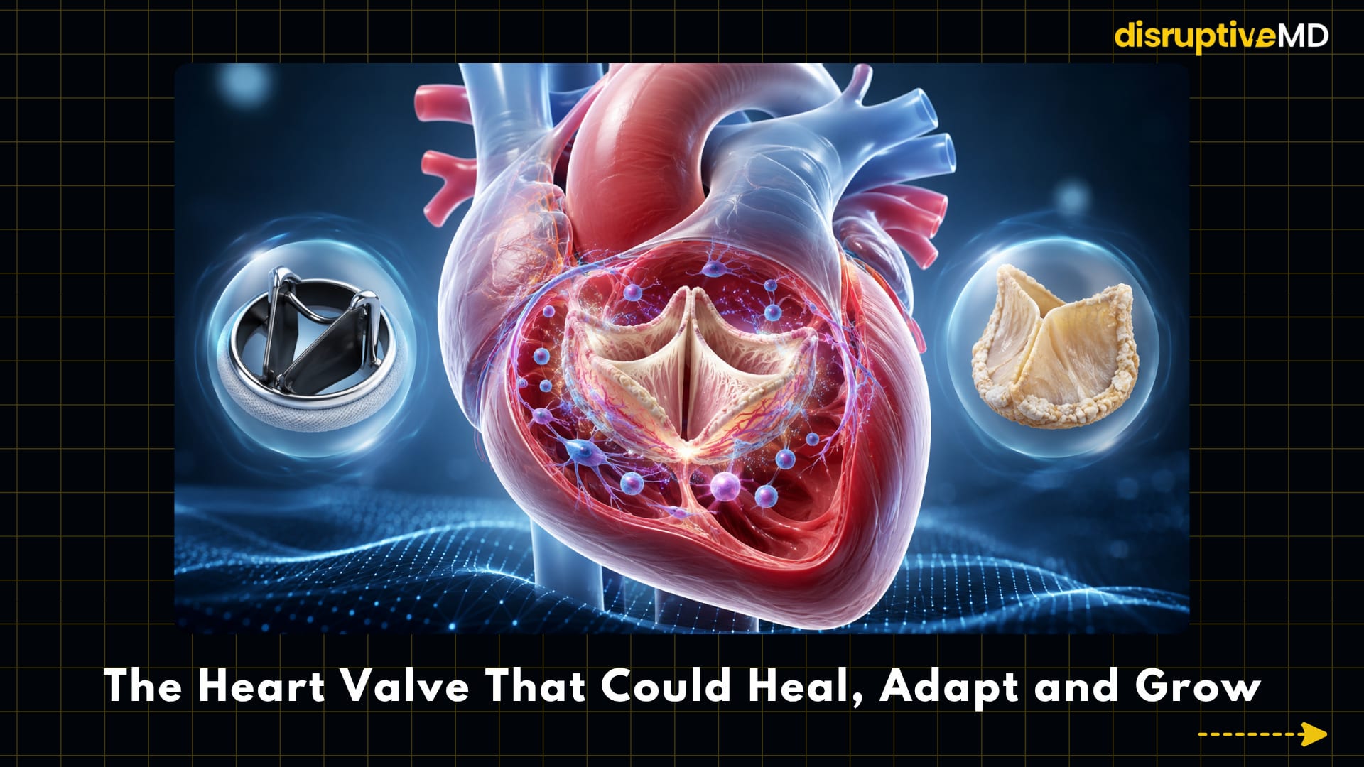
- Mechanical valves offer exceptional durability, but most patients require lifelong anticoagulation.
- Biological valves often avoid permanent anticoagulation, but they can stiffen, calcify and deteriorate over time.
- These valves could recruit cells, remodel their own structure, resist calcification and potentially grow as a child grows.
The Current Valve Compromise
Mechanical valves are manufactured from highly durable materials and can function for decades. Their artificial surfaces and flow patterns, however, can activate platelets and promote thrombosis. Lifelong anticoagulation lowers this risk but introduces another problem: bleeding.
Bioprosthetic valves behave more like natural tissue and are commonly used in both surgical and transcatheter valve replacement. They are less thrombogenic than mechanical valves, but they are not living structures. They cannot repair accumulated damage, respond normally to biological stress or grow with the patient.
Over time, biological valve leaflets may undergo:
- calcification
- collagen damage
- inflammation
- tearing or stiffening
- structural valve deterioration
- narrowing or leakage
This can eventually lead to another operation or a valve-in-valve procedure. Current guidelines therefore describe bioprosthetic durability as finite, with faster deterioration generally expected in younger patients.
Bioengineering asks a different question: What if the replacement valve were designed to repair and remodel instead of simply wearing out?
Why Engineering a Heart Valve Is So Difficult
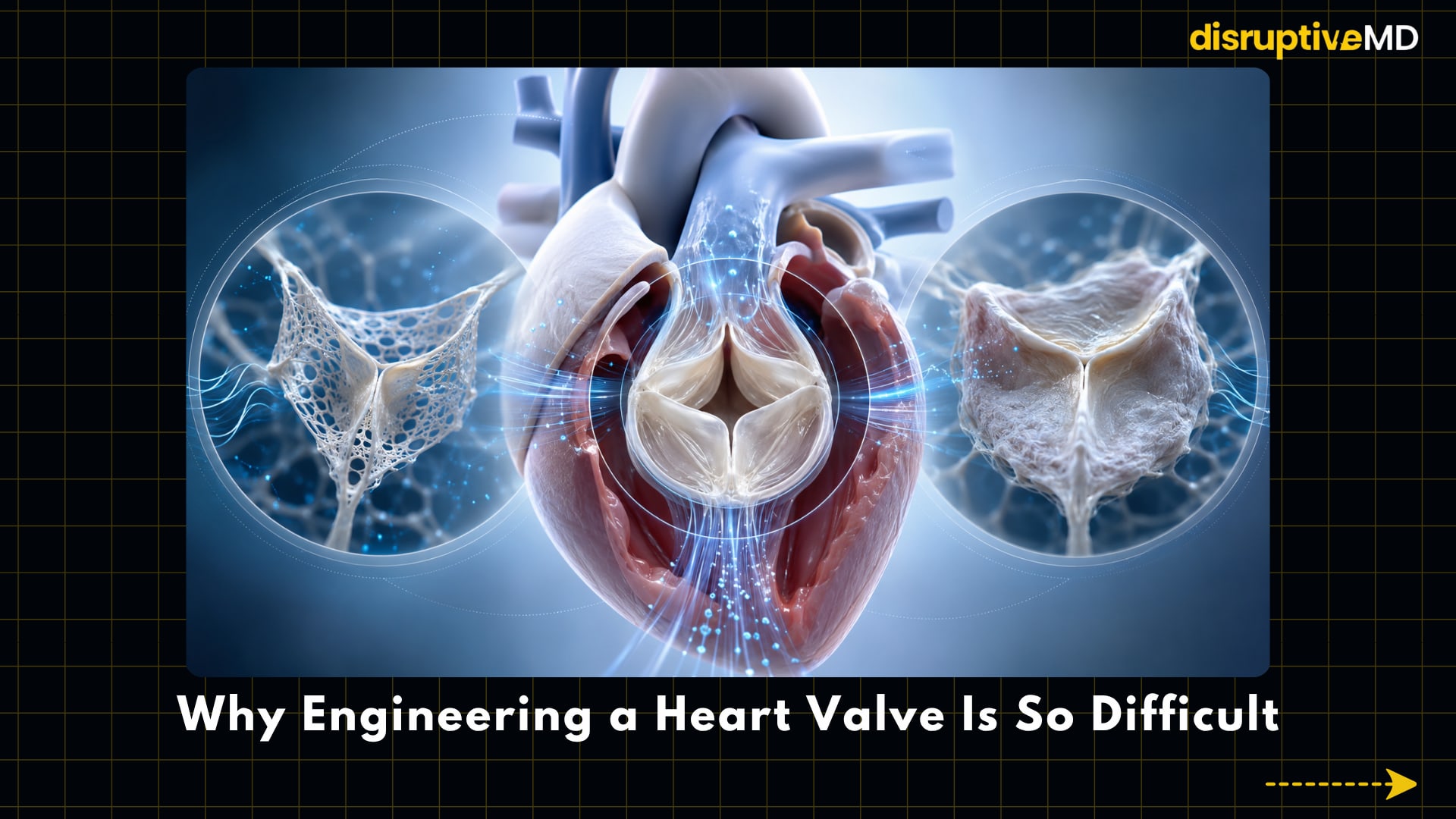
A heart valve may appear structurally simple, but it is one of the body’s most demanding mechanical systems.
Its thin leaflets must open fully, close precisely and prevent backward blood flow during every heartbeat. At the same time, the valve must remain flexible, resist clot formation, tolerate changing pressures and survive repeated loading for years.
A successful living valve must achieve several goals simultaneously:
- Function correctly from the moment it is implanted.
- Withstand the pressure and movement of circulating blood.
- Encourage healthy cell attachment.
- Avoid excessive inflammation and scar formation.
- Develop organised collagen and elastin.
- Resist thrombosis and calcification.
- Maintain its shape while its scaffold remodels.
- Potentially enlarge without becoming leaky or obstructed.
This balance is difficult. If tissue formation is too slow, the valve may weaken before the body can replace the scaffold. If healing is too aggressive, thick scar tissue may restrict leaflet movement.
The same biological response intended to regenerate the valve can therefore become the reason it fails.
Building a Living Valve
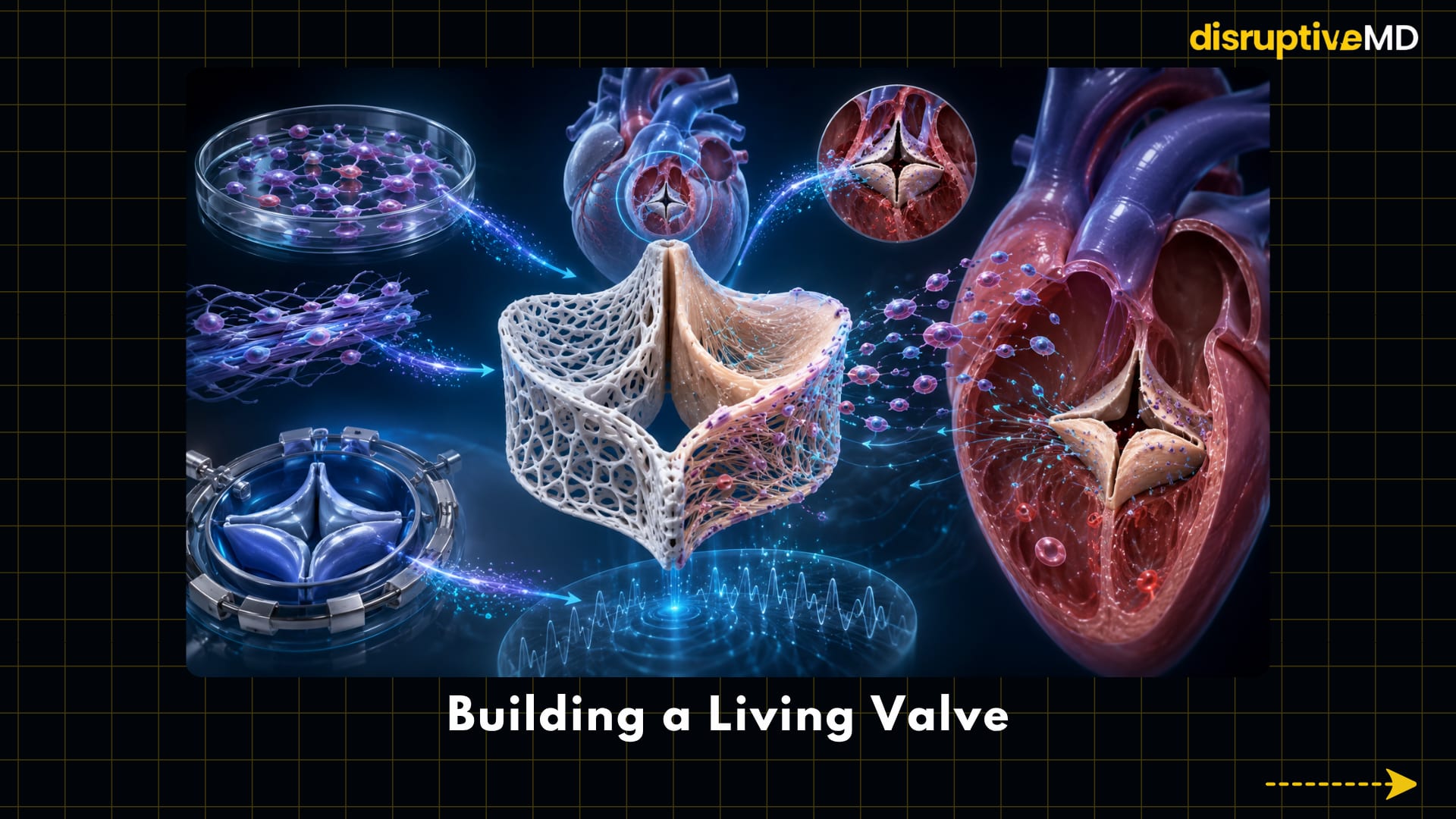
Tissue-engineered heart valves combine three major elements:
A scaffold provides the initial valve shape and mechanical support.
Cells may be added before implantation or recruited from the patient after implantation.
Biological and mechanical signals guide those cells to create organised, functional tissue.
The scaffold can be made from natural extracellular matrix, biodegradable polymers, hydrogels or combinations of synthetic and biological materials. It must be strong enough to function immediately but compatible enough to support long-term tissue development.
There are three major strategies being explored.
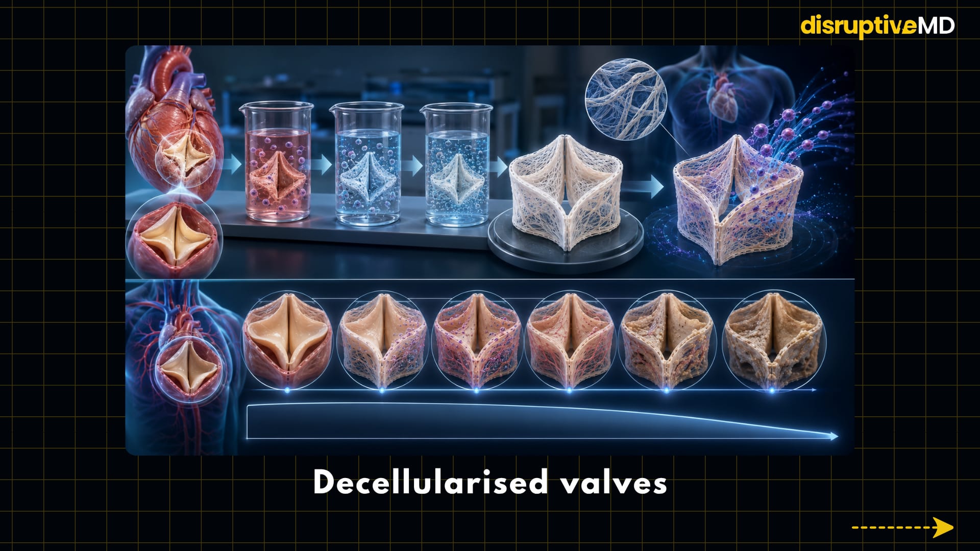
1. Decellularised valves
Decellularisation removes cells and cellular material from donor tissue while attempting to preserve its extracellular matrix.
This leaves behind a natural three-dimensional structure containing collagen, elastin and other components that originally supported the valve. The remaining scaffold may then be repopulated by the recipient’s cells.
Removing donor cells could reduce immune activation, but it does not automatically create a perfect valve. Processing must remove enough cellular material without damaging the matrix. Sterilisation, storage, donor variability, incomplete recellularisation and residual immunogenicity can all affect performance.
Decellularised pulmonary homografts already have meaningful clinical experience in selected congenital and pulmonary valve procedures. A 2026 report describing approximately 20 years of experience found favourable freedom from explantation, but also documented declining valve function over time. This shows both the promise and the remaining limitations of the approach.
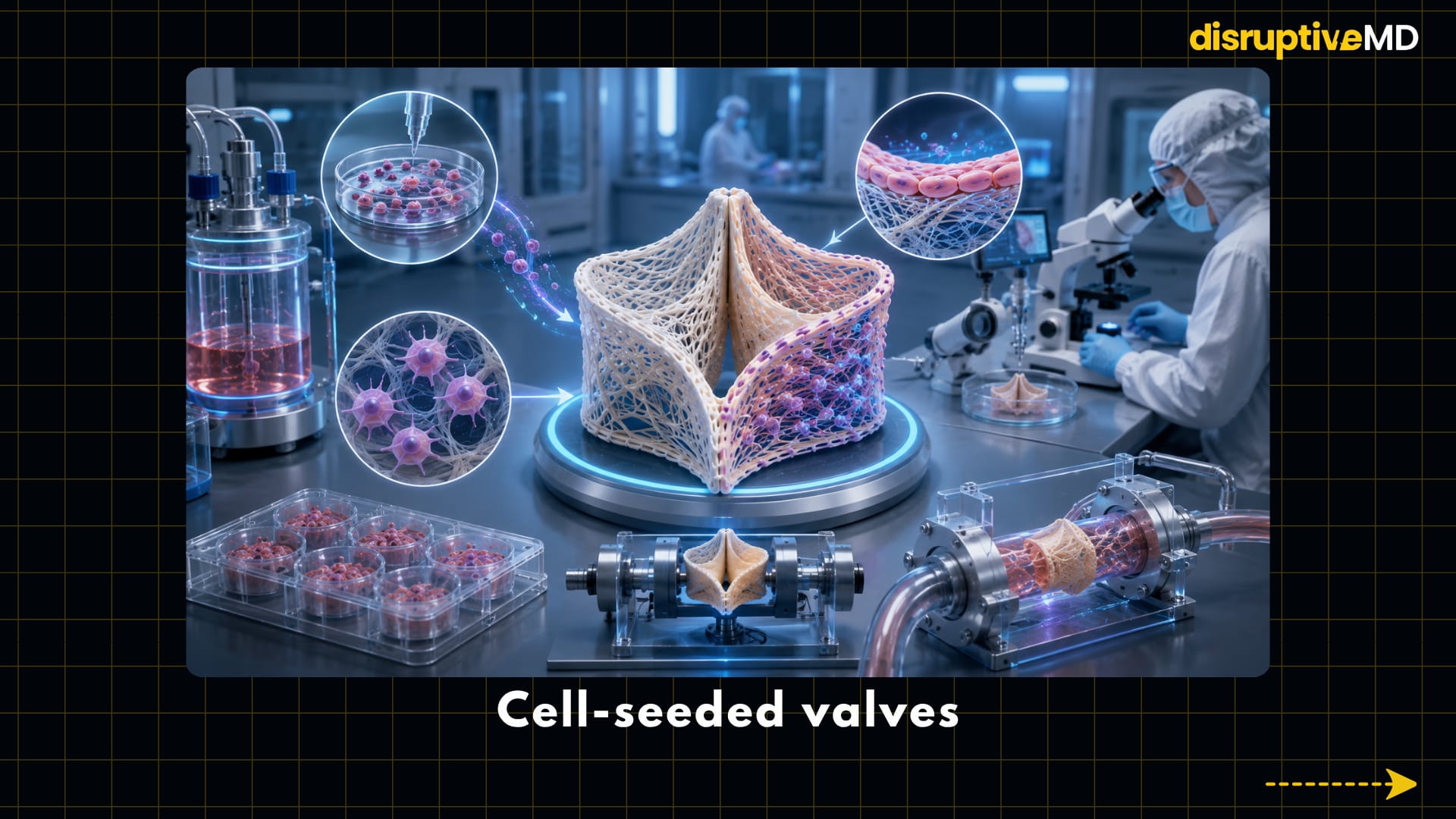
2. Cell-seeded valves
Another approach is to place patient-derived or laboratory-grown cells onto a scaffold before implantation.
Endothelial cells may help create a blood-compatible surface, while fibroblast-like or progenitor cells may produce extracellular matrix and support tissue repair.
Early clinical research has demonstrated that autologous cells can be used to reseed decellularised pulmonary valves. However, producing a personalised cell-seeded valve is complex. Cells must be collected, expanded, tested and distributed throughout the scaffold while maintaining sterility and consistent mechanical performance.
The implant must also work immediately. It cannot wait several months for the tissue to mature before beginning its job.
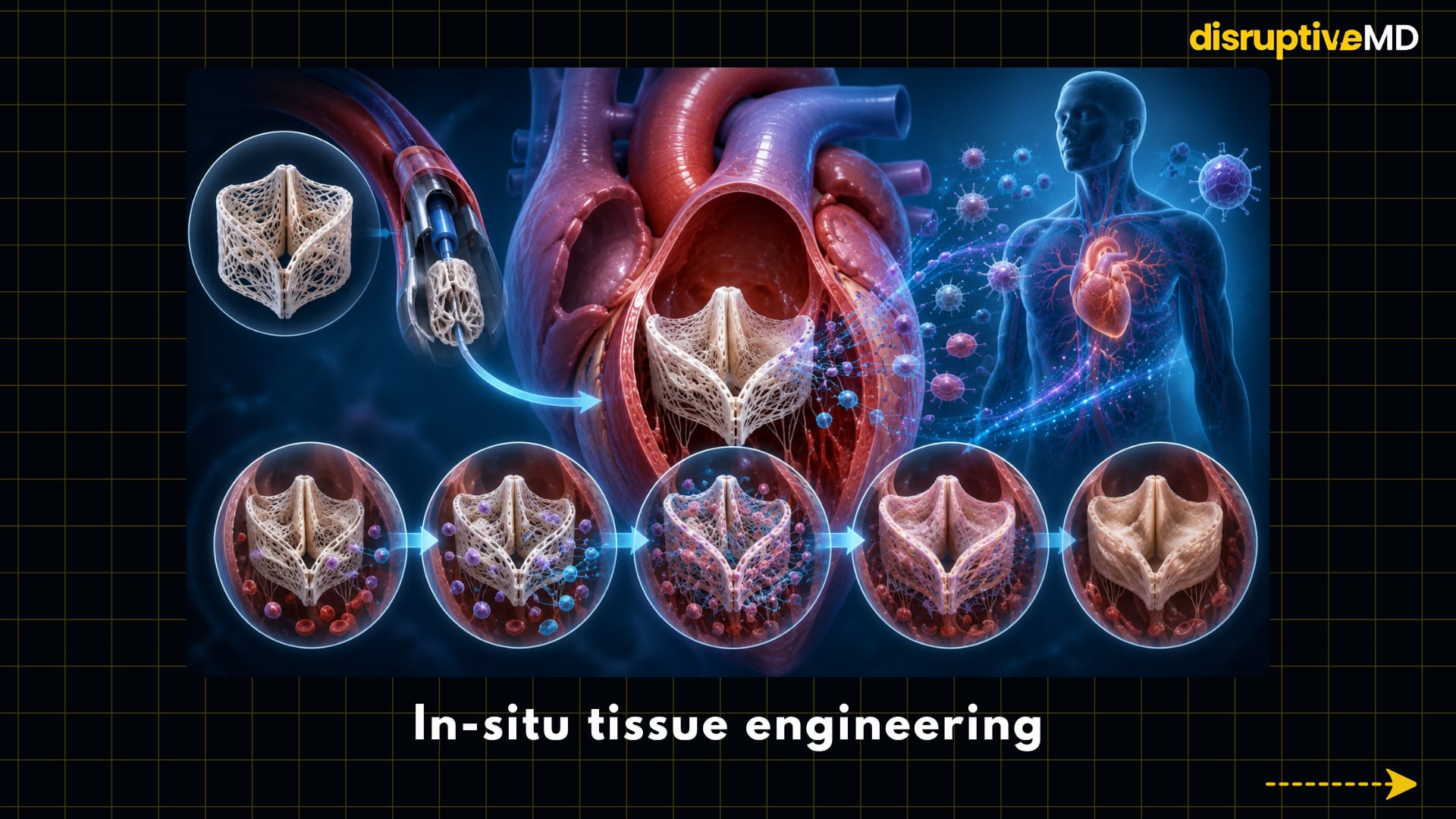
3. In-situ tissue engineering
In-situ tissue engineering removes some of the laboratory manufacturing burden.
A biodegradable, cell-free scaffold is implanted directly into the patient. Once inside the body, it recruits circulating and local cells. These cells gradually produce new tissue while the original scaffold breaks down.
In effect, the body becomes part of the manufacturing system.
Synthetic in-situ valves have maintained function for up to 12 months in preclinical sheep models, demonstrating that a bioresorbable scaffold can support tissue formation while continuing to open and close. However, controlling the quality, organisation and long-term stability of the regenerated tissue remains a major challenge.
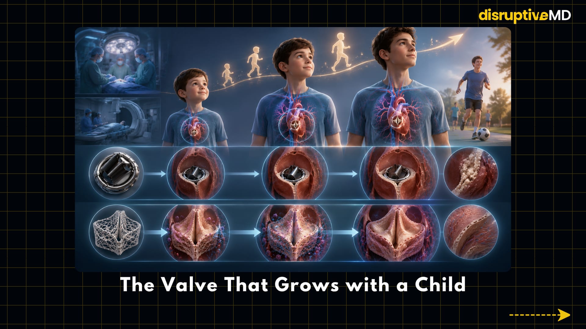
The Valve That Grows with a Child
The most compelling application may be paediatric heart surgery.
Children born with congenital heart disease can outgrow fixed-size prosthetic valves and conduits. Even a perfectly functioning implant may become too small as the heart and circulation enlarge.
This can lead to repeated catheter procedures or open-heart operations throughout childhood.
A genuinely growing valve would need to increase its dimensions through the formation of new living tissue while preserving normal leaflet movement, blood flow and structural strength.
Researchers have demonstrated remodelling and dimensional changes in experimental cardiovascular implants, but true functional growth remains difficult to prove. Enlargement alone is not enough. A valve must grow because it has produced new organised tissue—not because it has stretched, dilated or begun to fail.
A bioengineered valve that reliably grows with every child is therefore not yet available. But achieving it could fundamentally change the lifetime treatment pathway for congenital heart disease.
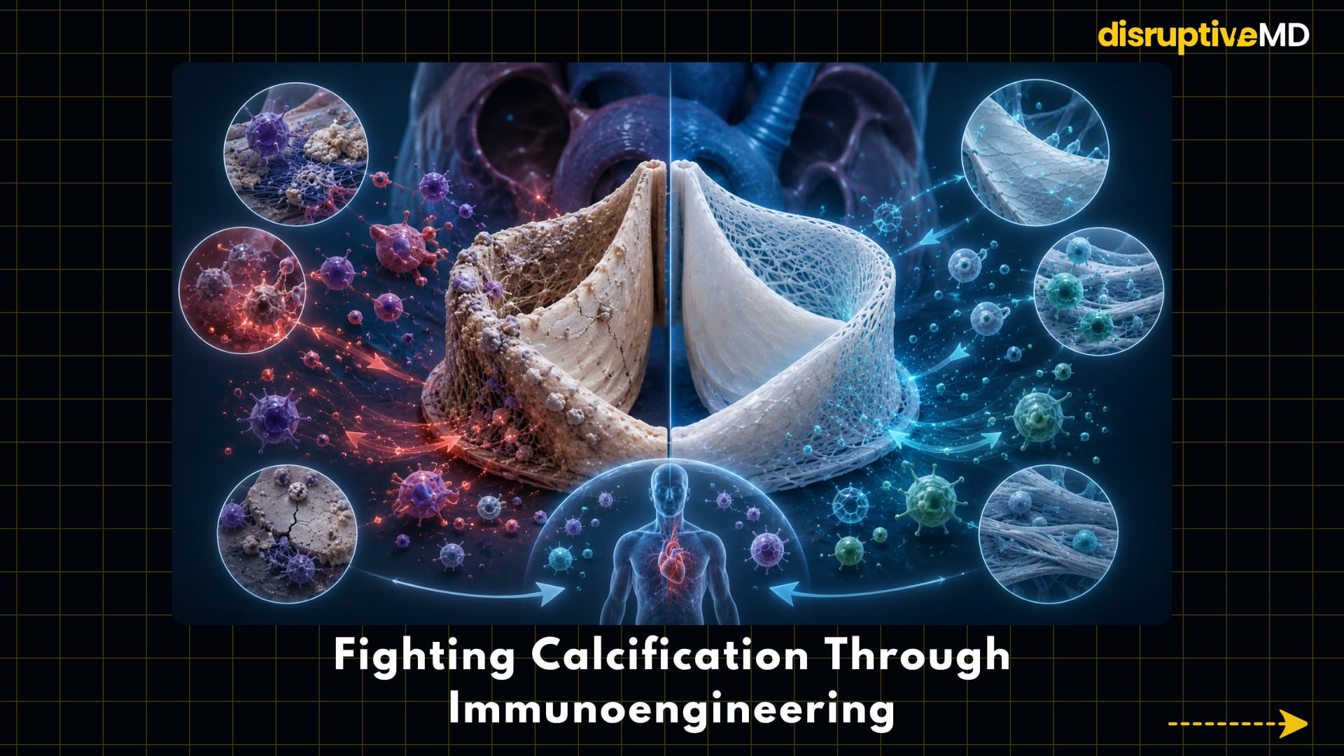
Fighting Calcification Through Immunoengineering
Calcification is not simply passive mineral accumulation. It can be influenced by mechanical stress, cell damage, inflammatory signalling, immune activity and the chemical treatment used to preserve biological tissue.
Future valves may use several strategies to reduce it:
- non-calcifying scaffold materials
- improved tissue preservation
- anti-inflammatory surfaces
- controlled scaffold degradation
- immune-modulating molecules
- endothelialised blood-contacting surfaces
- extracellular matrices designed for healthy remodelling
This is where tissue engineering increasingly overlaps with immunoengineering.
Rather than attempting to make the implant biologically invisible, researchers may design it to guide the immune response. Early immune cells could be encouraged to support constructive healing instead of prolonged inflammation, fibrosis or calcification.
The goal is not to eliminate the immune response. Healing requires immunity. The goal is to make the response regenerative rather than destructive.
Patient-Specific Valves and 3D Bioprinting
Valve anatomy differs between patients. Congenital abnormalities, calcification, previous surgery and changes in the surrounding heart can make standardised devices difficult to fit.
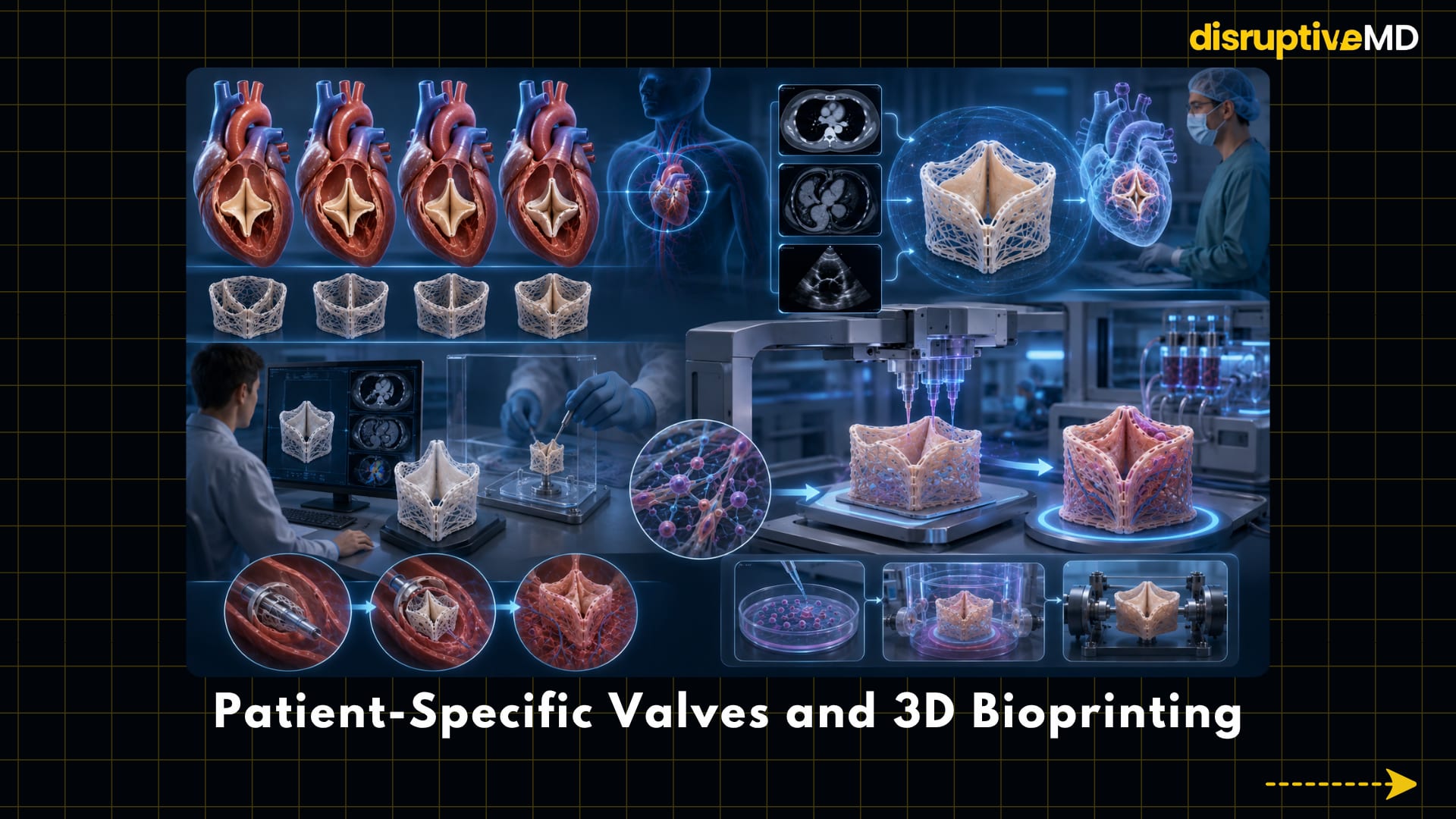
Three-dimensional imaging can already reconstruct individual valve anatomy from CT, MRI and echocardiography. These models can help clinicians study leaflet geometry, annular shape, calcification and the relationship between the valve and nearby structures.
3D printing can turn these datasets into physical models for procedural planning and device testing.
Bioprinting goes further by placing cells and biomaterials layer by layer to create tissue-like structures. In theory, it could produce valves with patient-specific geometry, spatially organised cell populations and different material properties across the leaflet.
However, printing a valve shape is not the same as creating a durable valve.
A clinically useful bioprinted valve must survive blood pressure, develop a non-thrombogenic surface, maintain microscopic tissue organisation and continue functioning through millions of cycles. Current cardiac bioprinting research is progressing rapidly, but fully bioprinted replacement valves are not part of routine clinical care.
Transcatheter Regeneration
The next major step may be to combine regenerative materials with minimally invasive delivery.
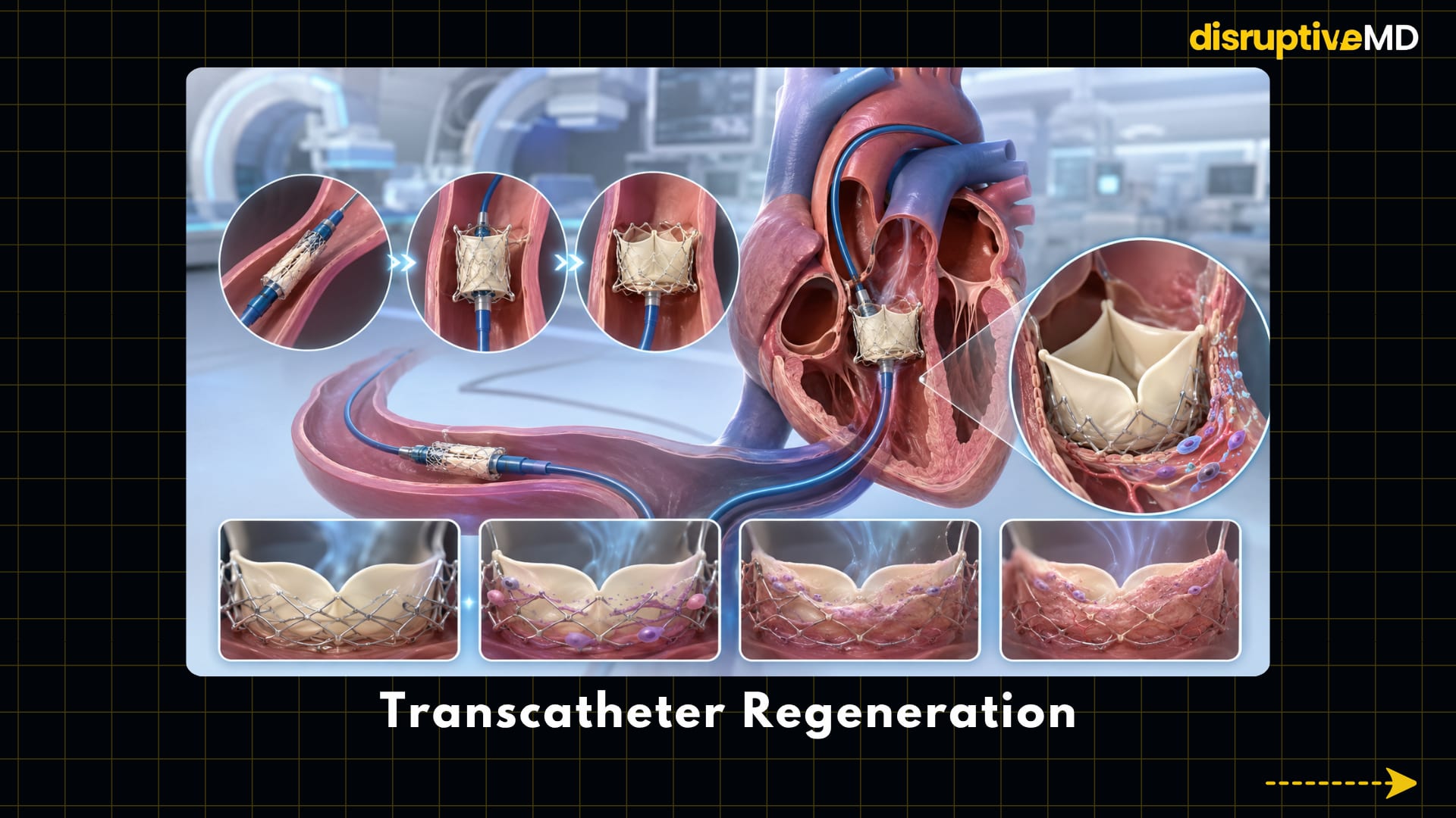
Transcatheter valve replacement has already changed the treatment of aortic valve disease by allowing a collapsible biological valve to be delivered through a catheter rather than through conventional open-heart surgery.
Bioengineered valves could eventually use a similar pathway.
A regenerative valve might be compressed onto a stent, delivered through a blood vessel and expanded inside the heart. After implantation, its scaffold could recruit cells and gradually remodel into living tissue.
But transcatheter delivery adds another engineering challenge. The valve must be thin and flexible enough to be compressed, strong enough to function immediately and biologically active enough to regenerate after deployment.
The processes of crimping and expansion must not damage the cells, scaffold or leaflet structure.
AI and the Digital Twin Valve
A bioengineered valve may behave differently in every patient because tissue growth is influenced by anatomy, blood flow, immune response and mechanical stress.
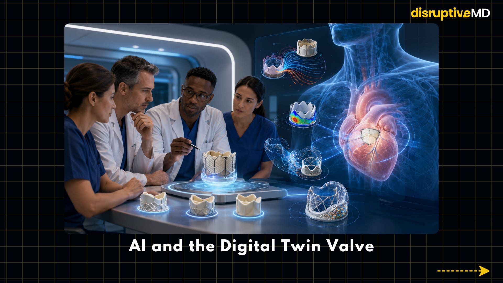
AI and computational modelling could help predict these interactions before implantation.
A patient-specific digital model could simulate:
- valve opening and closure
- pressure gradients
- leaflet stress
- blood-flow patterns
- leakage around the valve
- thrombotic flow zones
- scaffold degradation
- tissue growth and remodelling
- long-term fatigue
Patient-specific TAVR simulations have already shown how alternative deployment positions can influence leakage and valve behaviour. More advanced digital-twin systems are being investigated for structural-heart planning, but clinical validation and standardisation remain incomplete.
In the future, doctors may test several valve shapes, materials and implantation positions virtually before selecting the final design.
The digital twin would not replace the heart team. It would provide a personalised testing environment for decisions that currently rely heavily on population averages.
Fact Base
- Mechanical valves remain highly durable but generally require lifelong vitamin K antagonist anticoagulation.
- Bioprosthetic valves are widely used surgically and through transcatheter procedures, but they remain vulnerable to finite durability and structural deterioration.
- Decellularised pulmonary valves have been implanted clinically, particularly in congenital and right-sided heart procedures. Long-term studies show promising performance but not permanent freedom from degeneration.
- Cell-seeded valves have reached limited early clinical use, but manufacturing complexity restricts widespread adoption.
- Bioresorbable in-situ tissue-engineered valves have shown functional regeneration in large-animal studies, but dependable human outcomes have not yet been established.
- Patient-specific computational valve modelling is already technically possible and is being studied for procedural planning.
- 3D-printed anatomical models are clinically useful in selected planning applications, while fully living bioprinted replacement valves remain experimental.
- Reliable growth, lifelong durability and complete resistance to thrombosis or calcification have not yet been demonstrated in a routinely available bioengineered valve.
What Is Possible Today
Mechanical, surgical bioprosthetic and transcatheter tissue valves are established treatments.
Decellularised donor valves are used in selected pulmonary and congenital procedures.
Scaffold-based, cell-seeded and in-situ tissue-engineered valves are active areas of laboratory, animal and early translational research.
Patient-specific imaging and computational modelling are increasingly important in complex valve planning.
Bioengineered materials are also being developed to improve blood compatibility, flexibility and resistance to calcification.
What Is Not Fully Real Yet
There is currently no universally available living valve that reliably repairs itself for life.
There is no routinely implanted valve proven to grow normally with every child.
Fully 3D-bioprinted replacement valves are not standard clinical products.
Bioengineered valves have not eliminated thrombosis, calcification, infection or structural deterioration.
Digital twins cannot yet predict years of biological remodelling with complete accuracy.
Mechanical and conventional biological valves therefore remain essential. Bioengineered valves are not ready to replace every existing option.
Key Takeaway
Bioengineered heart valves represent a shift from replacement to regeneration.
The goal is no longer simply to manufacture a stronger artificial valve. It is to create a structure that functions immediately, communicates with the immune system, recruits the patient’s cells and gradually develops into living tissue.
The greatest opportunity may be in children, where growth could prevent repeated surgeries. But reaching that goal will require more than successful tissue formation. Researchers must prove that a regenerated valve can maintain its geometry, flexibility, blood compatibility and durability for decades.
The next generation of heart valves may combine biodegradable scaffolds, immunoengineering, patient-specific design, transcatheter delivery and digital simulation.
The future heart valve may not just open and close - it may heal, adapt and grow.
References
- Otto CM, et al. 2020 ACC/AHA Guideline for the Management of Patients With Valvular Heart Disease. Circulation. DOI: 10.1161/CIR.0000000000000923.
- Sarikouch S, et al. 20-Year Experience With Decellularized Pulmonary Homografts for Pulmonary Valve Replacement. European Journal of Cardio-Thoracic Surgery. 2026. DOI: 10.1093/ejcts/ezag087.
- Boethig D, et al. A European Study on Decellularized Homografts for Pulmonary Valve Replacement: Initial Results From the ESPOIR Trial.
- Cebotari S, et al. Clinical Application of Tissue Engineered Human Heart Valves Using Autologous Progenitor Cells. Circulation. 2006.
- Kluin J, et al. In Situ Heart Valve Tissue Engineering Using a Bioresorbable Elastomeric Implant: From Material Design to 12-Month Follow-Up in Sheep. Biomaterials. 2017.
- Bianchi M, et al. Patient-Specific Simulation of Transcatheter Aortic Valve Replacement: Impact of Deployment Options on Paravalvular Leakage. Biomechanics and Modeling in Mechanobiology. DOI: 10.1007/s10237-018-1094-8.
- Esmaeili A, et al. Advancements and Challenges in Tissue Engineering of Heart Valves: From Bioprinting to Clinical Applications. Polymer Advanced Technologies. 2025. DOI: 10.1002/pat.70226.