
What if ultrasound was no longer a machine you visit, but a patch you wear?
For decades, ultrasound has been something that happens during an appointment.
A clinician places a probe against the body, finds the correct acoustic window, captures images and removes the probe. The information can be incredibly valuable — but it is still largely a snapshot of what the body was doing at that particular moment.
Wearable ultrasound could change that model completely.
Instead of holding a probe for a few minutes, imagine placing a thin ultrasound patch on the skin and allowing it to watch what is happening underneath for hours.
Blood vessels could be followed as pressure changes.
Muscles could be observed while they contract.
The heart could potentially be monitored during exercise and recovery.
Bladder filling could be tracked automatically.
Selected fetal and blood-flow signals could be followed during pregnancy.
And rather than asking someone to remain still beside an ultrasound machine, the imaging system could begin moving with the patient.
Research prototypes have already demonstrated long-duration organ imaging, wearable cardiac imaging, deep-tissue monitoring in moving subjects, bladder-volume assessment and continuous cardiovascular measurements.
The real goal is not simply to make ultrasound smaller.
It is:
To turn ultrasound from a one-time imaging test into a continuous internal health monitor.
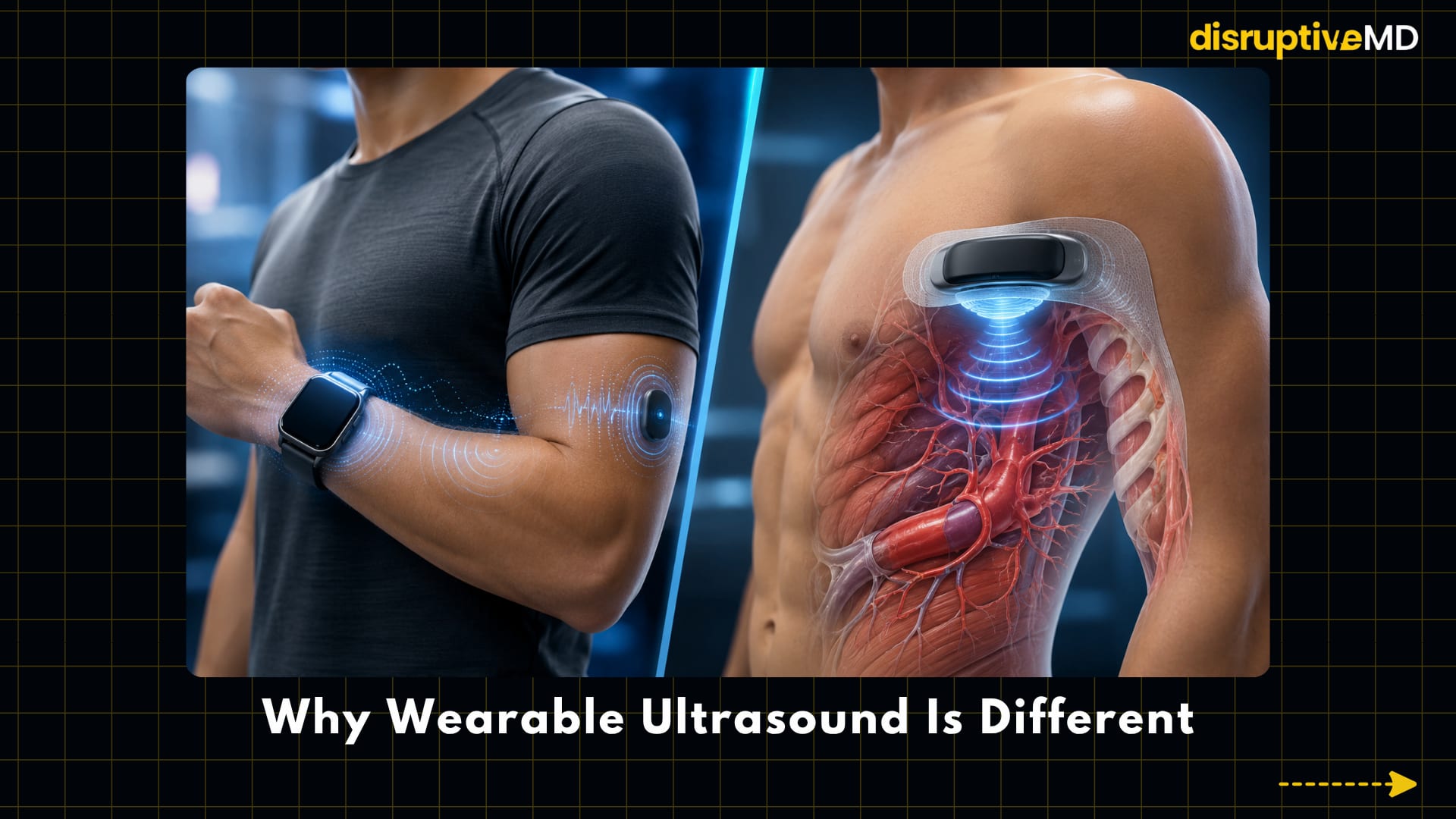





- Smartwatches and patches can track heart rate, ECG signals, oxygen saturation, temperature, movement, sleep and physical activity.
- Sound waves can interrogate tissues beneath the skin and provide information about anatomy, movement, blood vessels and physiological changes without using ionising radiation.
- Wearable ultrasound asks: What can we continuously observe on the inside?
WHY WEARABLE ULTRASOUND IS DIFFERENT
Modern wearables are already remarkably good at measuring the surface of the body.
Smartwatches and patches can track heart rate, ECG signals, oxygen saturation, temperature, movement, sleep and physical activity.
But most of those signals originate at, or are inferred from, the body’s surface.
Ultrasound can go deeper.
Sound waves can interrogate tissues beneath the skin and provide information about anatomy, movement, blood vessels and physiological changes without using ionising radiation.
Wearable ultrasound therefore introduces a fundamentally different type of wearable.
The smartwatch asks:
What signals can we measure from the outside?
Wearable ultrasound asks:
What can we continuously observe on the inside?
That distinction is why the technology could become so important.
1. FROM HANDHELD PROBE TO SMART PATCH
Conventional diagnostic ultrasound remains highly dependent on the operator.
The probe must be positioned correctly. Its angle matters. Contact with the skin matters. Even small changes in orientation can dramatically change the image.
Making ultrasound wearable therefore requires more than simply shrinking a probe.
The transducer needs to conform to the body, remain coupled to the skin and maintain a useful acoustic window as the person moves.
One important breakthrough came with bioadhesive ultrasound, where a thin ultrasound device could remain attached to the skin for prolonged imaging.
A 2022 study demonstrated continuous imaging for 48 hours, including imaging of blood vessels, muscle, the heart, gastrointestinal tract and diaphragm.
Later systems pushed the idea further by integrating flexible electronics, ultrasound arrays, wireless communication and machine-learning algorithms into increasingly autonomous devices.
A fully integrated wearable ultrasound system reported in Nature Biotechnology could track physiological signals from tissues as deep as 164 mm and monitor parameters including central blood pressure, heart rate and cardiac output in moving participants for periods of up to 12 hours.
That changes what ultrasound could eventually become. Instead of asking:
“What does this organ look like right now?”
medicine may increasingly be able to ask:
“How has this organ been behaving for the last several hours?”
2. CONTINUOUS HEART MONITORING
The heart is an obvious target for wearable ultrasound.
A smartwatch can measure pulse.
An ECG can measure the heart’s electrical activity.
But neither directly shows the heart muscle contracting or its chambers filling and emptying.
Ultrasound can.
A wearable cardiac ultrasound system reported in Nature demonstrated continuous, real-time imaging of cardiac function while attached to the chest. The device could examine the left ventricle during different conditions, including movement.
The researchers also developed a deep-learning system capable of extracting left-ventricular volume from the continuous images and generating measurements including:
- Stroke volume
- Cardiac output
- Ejection fraction
This points toward a completely different form of cardiovascular monitoring.
Instead of seeing cardiac function only during an echocardiogram appointment, future systems could potentially examine how the heart responds to:
exercise, stress, rehabilitation, medication, recovery or worsening disease.
For heart failure, critical care and post-procedure monitoring, that could become particularly powerful.
The wearable would no longer simply tell us how fast the heart is beating.
It could begin telling us how the heart is mechanically performing.
3. BLOOD PRESSURE WITHOUT JUST THE CUFF
Blood pressure monitoring could also become much deeper.
Traditional arm cuffs provide intermittent measurements.
Continuous arterial pressure monitoring in critically ill patients can require an invasive arterial catheter.
Wearable ultrasound introduces another possibility:
watch the artery itself.
Ultrasound can track arterial wall motion and pulsatile changes associated with each heartbeat.
This concept has progressed considerably beyond early engineering demonstrations.
A wearable ultrasound blood-pressure sensor has been tested during daily activities and in clinical environments including outpatient care, cardiac catheterisation and intensive care. The researchers reported that the device’s design improved reliability and met demanding clinical performance requirements in their validation studies.
That does not mean blood-pressure cuffs are disappearing.
But it suggests that cardiovascular wearables may eventually measure more than pulse rate and skin-level optical signals.
They may continuously interrogate the mechanical behaviour of blood vessels underneath the skin.
4. MUSCLE COULD BECOME A LIVE BIOMECHANICAL SIGNAL
Sports medicine and rehabilitation may be one of the most interesting applications.
Today we can measure movement with accelerometers.
We can measure external force with force plates.
We can estimate electrical muscle activation using surface electromyography.
But none of those directly show what the muscle itself is doing beneath the skin.
Ultrasound can.
Wearable ultrasound could potentially track:
- Muscle contraction
- Muscle thickness
- Changes in architecture
- Tissue deformation
- Tendon movement
- Tissue stiffness
- Recovery after injury
Stretchable ultrasound arrays have already been demonstrated for three-dimensional mapping of deep-tissue mechanical properties at depths of up to approximately 4 cm. Researchers used the system to investigate muscle changes and monitor recovery during physiotherapy.
That creates a fascinating possibility for rehabilitation.
Imagine an athlete performing a strengthening exercise while an ultrasound patch observes how the muscle behaves through each repetition.
A rehabilitation programme might eventually measure not only:
“How much force can you produce?”
but also:
“How is the tissue producing that force?”
For return-to-sport testing, sarcopenia, neuromuscular disease and post-operative rehabilitation, that additional layer of information could be extremely valuable.
5. PREGNANCY MONITORING BEYOND THE OCCASIONAL SCAN
Pregnancy may demonstrate the difference between snapshot imaging and longitudinal imaging better than almost any other application.
Conventional prenatal ultrasound provides rich anatomical and physiological information, but most examinations represent relatively short windows of observation.
Wearable ultrasound research is beginning to investigate what happens when those windows become much longer.
In 2026, researchers reported a wearable ultrasound patch developed for continuous and autonomous fetal monitoring.
The system could acquire fetal anatomical information and blood-flow velocities and showed good agreement with handheld clinical ultrasound measurements across 62 pregnancies.
This is particularly interesting for research involving high-risk pregnancies.
Instead of assessing selected fetal or circulatory parameters only during scheduled examinations, wearable systems could eventually allow certain measurements to be followed over longer periods.
But the distinction between research and routine care is essential.
Wearable fetal ultrasound is not currently a replacement for conventional prenatal ultrasound, specialist fetal assessment or established pregnancy monitoring.
What the research demonstrates is something different:
Continuous wearable fetal ultrasound is becoming technically possible.
6. A PATCH THAT KNOWS WHEN THE BLADDER IS FULL
Not every disruptive medical technology has to solve a dramatic problem.
Sometimes the most useful applications are extremely practical.
Bladder monitoring is one example.
Patients with urinary retention, neurogenic bladder, neurological injury or certain post-operative conditions may require repeated assessment of bladder filling.
A wearable ultrasound patch could potentially turn the bladder into something that can be monitored continuously rather than repeatedly scanned manually.
A conformable phased-array ultrasound patch has already been tested in a pilot clinical study for bladder-volume assessment.
The researchers reported bladder-volume measurements comparable with standard clinical ultrasound, while the patch avoided the need for repeatedly translating or rotating a handheld probe.
In the future, such a system could potentially work more like an internal gauge:
Bladder fills → ultrasound detects the change → system estimates volume → patient or clinician receives an alert.
That is a very different role for ultrasound.
It stops behaving purely like an imaging examination and starts behaving like a continuous physiological sensor.
7. COULD IT MONITOR THE LUNGS AND FLUID STATUS?
Ultrasound can already reveal certain patterns associated with fluid accumulation and changes at tissue interfaces.
That makes wearable monitoring of selected lung or fluid-related signals an intriguing possibility.
One future application could be monitoring patients whose fluid status changes over time.
Instead of waiting for symptoms to worsen or relying exclusively on weight, vital signs and occasional examinations, strategically positioned ultrasound sensors could potentially detect changing internal patterns.
But this remains more challenging than simply attaching a patch.
The lungs contain air, which strongly affects ultrasound propagation. Breathing moves anatomical structures. Body position changes the acoustic window. A patch may remain in exactly the same place on the skin while the underlying anatomy shifts.
So the future is unlikely to be:
“Wear a patch and continuously image every part of the lungs perfectly.”
A more realistic goal may be:
Monitor carefully selected ultrasound-visible features repeatedly and detect meaningful changes from the patient’s baseline.
That could still be clinically powerful.
8. AI COULD BECOME THE SECOND HALF OF THE TECHNOLOGY
Making ultrasound wearable creates another problem.
Too much data.
A conventional ultrasound examination may last minutes.
A wearable system could record for hours.
No clinician wants to manually review an eight-hour ultrasound recording frame by frame.
This is where artificial intelligence may become essential.
Machine-learning algorithms are already being incorporated into wearable ultrasound systems to track moving tissue targets and assist interpretation.
In wearable cardiac imaging, deep learning has already been used to automatically extract ventricular volume and calculate physiological measurements from continuous image recordings.
The future wearable ultrasound system may therefore not simply send a gigantic video file to a doctor.
Instead, software could potentially:
- Identify relevant anatomy
- Track structures as they move
- Remove or reduce motion artefacts
- Measure changes automatically
- Compare signals with previous recordings
- Detect unusual patterns
- Highlight important time periods for clinician review
The clinician might not need eight hours of ultrasound video.
The system could instead say:
“Cardiac output changed significantly during this period.”
Or:
“Bladder volume has crossed the programmed threshold.”
Or:
“Muscle mechanics during rehabilitation have changed compared with the previous session.”
That may be the combination that makes wearable ultrasound truly scalable:
continuous imaging + automated interpretation
9. FROM MONITORING TO CLOSED-LOOP MEDICINE
The next step goes beyond imaging.
What if ultrasound eventually became part of a system that can both observe and influence treatment?
The architecture could look like this:
Monitor tissue → detect change → adjust therapy → monitor the response
A rehabilitation device could potentially modify assistance according to muscle behaviour.
A cardiovascular monitoring platform could change surveillance intensity when haemodynamic patterns deteriorate.
Future drug-delivery systems, neurostimulation systems or smart implants might use internal ultrasound measurements as one of several feedback signals.
This is still largely a future concept rather than established clinical practice.
But it reflects a much larger direction in medicine.
Sensors are becoming continuous.
Algorithms are becoming predictive.
Therapies are becoming increasingly programmable.
Wearable ultrasound could add something crucial to that ecosystem:
continuous information from deep inside the body.
10. HOSPITAL-AT-HOME COULD GAIN AN INTERNAL WINDOW
Remote patient monitoring currently relies heavily on surface measurements.
Heart rate.
ECG.
Temperature.
Oxygen saturation.
Respiratory rate.
Movement.
Those signals are useful, but they reveal only part of the patient’s physiology.
Wearable ultrasound could potentially add another layer:
internal mechanical and anatomical information.
A patient recovering at home might one day wear an ultrasound device capable of following selected cardiac, vascular, muscular or organ-related measurements.
Instead of repeatedly bringing the patient to the imaging machine, certain forms of imaging could begin travelling with the patient.
That could become particularly relevant as hospital-at-home and remote-monitoring models expand.
The change would be subtle but profound.
Remote medicine would no longer monitor only what can be measured from the surface.
It could begin looking underneath it.
WHAT IS REAL TODAY?
Wearable ultrasound is no longer merely a futuristic concept.
What is already real in research
Researchers have demonstrated:
- Bioadhesive ultrasound patches capable of prolonged imaging of multiple internal structures.
- Wearable cardiac ultrasound capable of continuous real-time assessment of cardiac function.
- Fully integrated systems capable of monitoring deep tissues in moving subjects for hours.
- Wearable ultrasound approaches for continuous blood-pressure monitoring with clinical validation.
- Stretchable ultrasound arrays capable of mapping deep-tissue mechanical properties and monitoring muscle recovery.
- Conformable ultrasound patches for bladder-volume monitoring.
- Wearable fetal ultrasound research capable of acquiring anatomical information and blood-flow measurements during pregnancy.
These are substantial advances.
But they should not be confused with widespread everyday clinical availability.
What is not fully real yet
We do not yet have:
- A universal consumer ultrasound patch for everyday diagnosis
- Fully autonomous whole-body ultrasound interpretation
- Perfect image quality during every type of movement
- Reliable imaging of every organ from a fixed wearable position
- Comfortable long-term use in every patient
- Universal AI interpretation of ultrasound
- Routine continuous ultrasound monitoring for every disease
- Replacement of trained sonographers, radiologists or cardiologists
- A single patch capable of monitoring everything happening inside the body
Wearable ultrasound remains an emerging medical technology rather than a universal replacement for conventional diagnostic ultrasound.
THE BIGGEST CHALLENGES
Making ultrasound flexible is only one part of the problem.
The device must also maintain stable acoustic coupling with the skin.
It must stay positioned over the correct anatomy.
It must tolerate sweat, movement and changes in posture.
The electronics must become small enough for comfortable wear.
Power consumption needs to remain low.
Heat generation needs to be controlled.
Large quantities of ultrasound data need to be processed, stored or transmitted efficiently.
And long-term wear has to remain comfortable enough that patients will actually use the device.
Motion is particularly difficult.
The patch may remain attached perfectly while the organ underneath it moves because of breathing, exercise or changes in body position.
The system therefore needs to distinguish between:
a real physiological change
and
a change caused simply by the anatomy moving relative to the sensor.
Machine learning may help solve part of this problem, and integrated systems have already demonstrated automated tracking of moving tissue targets.
But engineering performance alone will not determine whether wearable ultrasound succeeds.
The technology must eventually demonstrate that continuous internal monitoring provides information that genuinely improves clinical decisions and patient outcomes.
THE BIGGER PICTURE
The smartwatch revolution showed that important health measurements do not always need to happen inside hospitals.
Wearable ultrasound could push that idea one layer deeper.
A patch on the chest could observe the heart.
A patch over an artery could follow vascular behaviour.
A patch over a muscle could examine tissue mechanics during rehabilitation.
A patch over the bladder could monitor filling.
Selected pregnancy patches could follow fetal and circulatory signals.
And AI could potentially transform hours of imaging into a small number of clinically meaningful measurements.
The important shift is therefore not simply:
large ultrasound machine → smaller ultrasound machine.
It is:
episodic imaging → continuous internal monitoring
Smartwatches showed us how valuable continuous physiological data could become.
Wearable ultrasound could extend that philosophy beneath the skin.
The next generation of wearables may not just count steps — they may look beneath the skin.
FACT BASE
Wearable ultrasound has progressed from early flexible-transducer experiments into increasingly integrated systems capable of prolonged and autonomous physiological monitoring.
A bioadhesive ultrasound platform demonstrated continuous imaging for up to 48 hours across several anatomical targets, providing an important proof of concept that ultrasound transducers could remain attached to the body for extended monitoring rather than being manually held in position.
Wearable cardiac imaging subsequently demonstrated continuous assessment of the heart using a skin-conformal ultrasound device. Deep-learning analysis was used to extract left-ventricular volume and derive stroke volume, cardiac output and ejection fraction from continuous recordings.
A fully integrated ultrasonic-system-on-patch combined a wearable transducer, miniaturised electronics, wireless communication and machine learning. The system tracked physiological signals from tissues as deep as 164 mm and monitored variables including central blood pressure, heart rate and cardiac output for up to 12 hours in mobile participants.
Wearable ultrasound has also expanded into organ-specific monitoring. A conformable phased-array bladder patch demonstrated volumetric bladder monitoring in a pilot clinical study, with estimated volumes comparable to conventional clinical ultrasound measurements. In musculoskeletal applications, stretchable ultrasonic arrays have demonstrated three-dimensional mapping of deep-tissue stiffness and were used to investigate muscle microstructural damage and recovery during physiotherapy.
Cardiovascular translation has progressed further with clinical evaluation of wearable ultrasound blood-pressure monitoring across daily activities, outpatient care, cardiac catheterisation and intensive care environments.
More recently, a 2026 study demonstrated a wearable fetal ultrasound patch capable of continuous and autonomous monitoring. The system acquired fetal anatomical structures and blood-flow velocities and was compared with handheld clinical ultrasound across 62 pregnancies.
Together, these studies establish that wearable ultrasound is technically capable of generating meaningful deep-tissue information for prolonged periods.
However, the field remains limited by stable acoustic coupling, motion artefacts, power requirements, electronics miniaturisation, heat management, wireless data transfer, automated interpretation, comfort, safety, clinical validation and integration into medical workflows.
The evidence therefore supports wearable ultrasound as a rapidly advancing research and emerging clinical platform, but not yet as a general replacement for standard diagnostic ultrasound.
REFERENCES
- Wang C, Chen X, Wang L, et al. Bioadhesive ultrasound for long-term continuous imaging of diverse organs. Science. 2022;377(6605):517–523. DOI: 10.1126/science.abo2542
- Hu H, Huang H, Li M, et al. A wearable cardiac ultrasound imager. Nature. 2023;613:667–675. DOI: 10.1038/s41586-022-05498-z
- Lin M, Zhang Z, Gao X, et al. A fully integrated wearable ultrasound system to monitor deep tissues in moving subjects. Nature Biotechnology. 2024;42:448–457. DOI: 10.1038/s41587-023-01800-0
- Zhang L, Marcus C, Lin D, et al. A conformable phased-array ultrasound patch for bladder volume monitoring. Nature Electronics. 2024;7:77–90. DOI: 10.1038/s41928-023-01068-x
- Hu H, Ma Y, Gao X, et al. Stretchable ultrasonic arrays for the three-dimensional mapping of the modulus of deep tissue. Nature Biomedical Engineering. 2023;7:1321–1334. DOI: 10.1038/s41551-023-01038-w
- Zhou S, et al. Clinical validation of a wearable ultrasound sensor of blood pressure. Nature Biomedical Engineering. 2025;9:865–881. DOI: 10.1038/s41551-024-01279-3
- Park G, Bian Y, Huang H, et al. Fetal monitoring for high-risk pregnancies using a wearable ultrasound patch. Nature Biotechnology. Published online May 26, 2026. DOI: 10.1038/s41587-026-03140-1