
What if artery plaque could be targeted from inside the bloodstream before it causes a heart attack or stroke?
For decades, atherosclerosis has often been described like a plumbing problem.
Cholesterol builds up.
The artery becomes narrow.
Blood flow is restricted.
The vessel eventually needs to be opened.
That description is easy to understand, but it is incomplete.
An atherosclerotic plaque is not simply a lump of fat sitting inside a blood vessel. It is a biologically active structure developing within the artery wall. It can contain cholesterol, immune cells, dead cells, scar-like tissue, calcium, tiny blood vessels and inflammatory signals.
Some plaques slowly become large and obstruct blood flow.
Others may cause little narrowing but remain highly inflamed, fragile and vulnerable to rupture. When the surface of such a plaque breaks, the body can respond by forming a blood clot. That clot, not necessarily the plaque’s physical size, may suddenly block blood flow and trigger a heart attack or stroke.
This changes the question completely.
The future of atherosclerosis treatment may not be about mechanically removing every visible deposit. It may be about changing the biology of the plaque before it becomes dangerous.
That is where nanotechnology enters the artery.
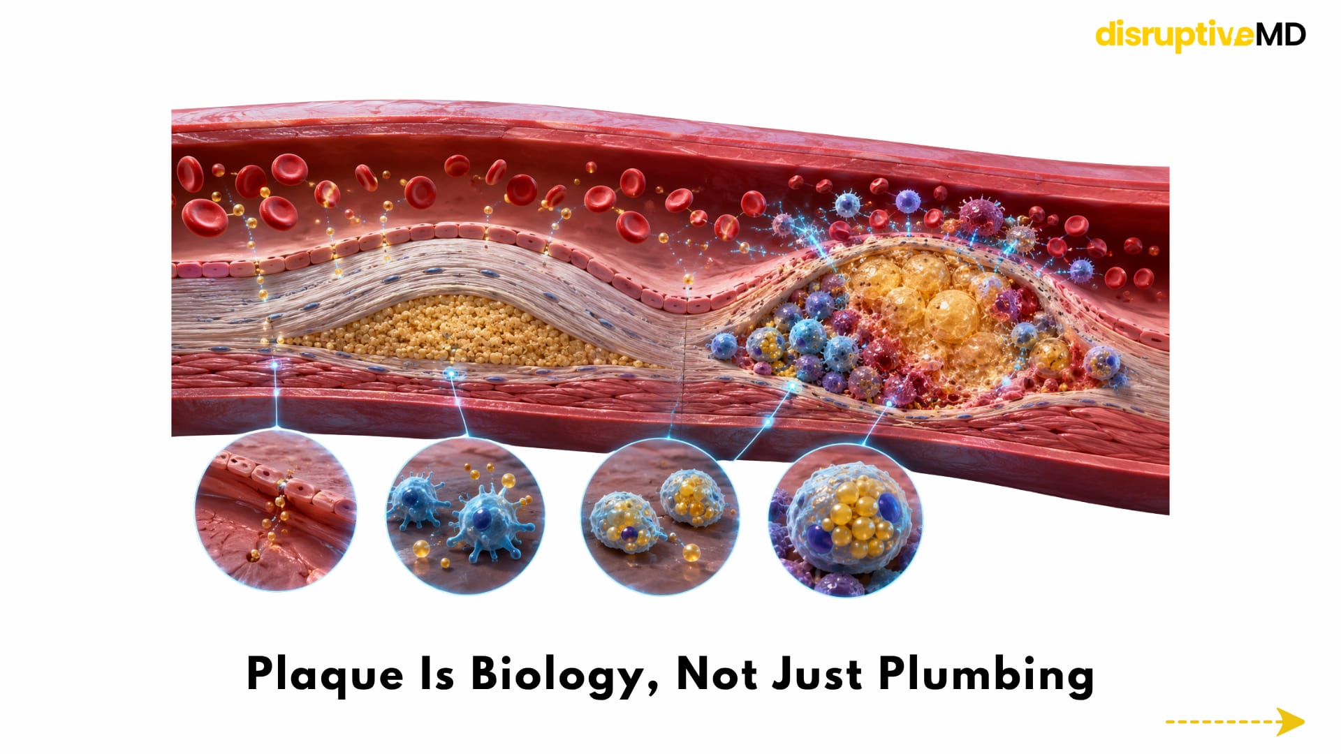
- Atherosclerosis begins when cholesterol-containing particles enter and become retained within the artery wall.
- As they become overloaded with lipids, many macrophages transform into foam cells.
- This is why the plaque producing the greatest narrowing is not automatically the plaque most likely to rupture.
Plaque Is Biology, Not Just Plumbing
Atherosclerosis begins when cholesterol-containing particles enter and become retained within the artery wall.
The artery responds as though it has been injured.
Immune cells are recruited. Inflammatory signals increase. Macrophages, cells that normally help remove harmful material and cellular debris, begin consuming modified cholesterol particles.
As they become overloaded with lipids, many macrophages transform into foam cells.
Some eventually die.
In a healthy healing response, dead cells should be rapidly recognised, engulfed and removed. Inside an advanced plaque, however, this clearance process can fail. Dead cells and extracellular lipids accumulate, contributing to the development of a soft, inflammatory necrotic core.
At the same time, the plaque attempts to isolate this dangerous material beneath a fibrous cap.
If that protective cap remains strong, the plaque may stay relatively stable. If inflammation, tissue-degrading enzymes and cellular death weaken it, the plaque can become more vulnerable to rupture.
This is why the plaque producing the greatest narrowing is not automatically the plaque most likely to rupture.
A moderately narrowed artery may contain an inflamed, lipid-rich lesion with a fragile surface. Another artery may contain a larger but more fibrotic and stable plaque.
Nanomedicine is attractive because nanoparticles can potentially be designed to recognise these biological differences, and not merely measure how narrow the artery has become.
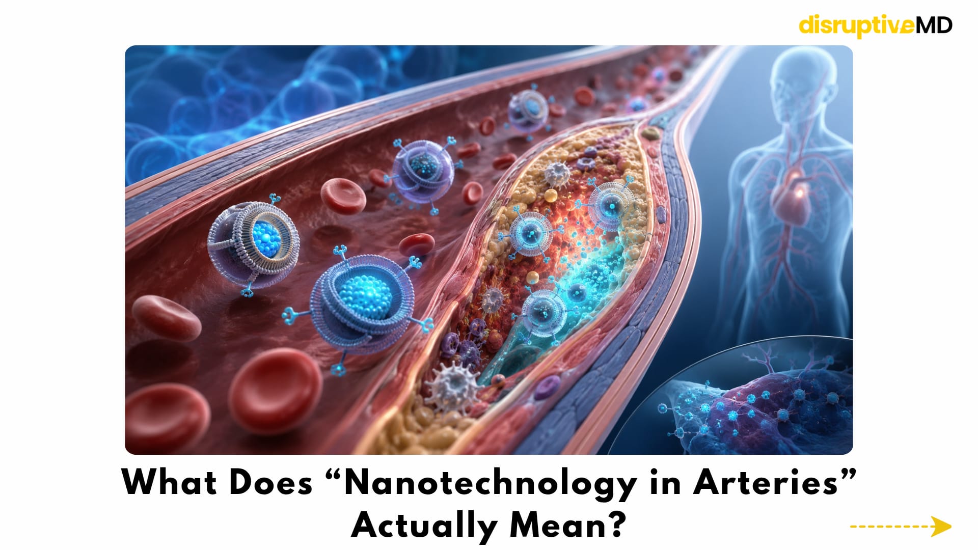
What Does “Nanotechnology in Arteries” Actually Mean?
Nanotechnology does not necessarily mean microscopic robots carrying miniature drills.
In medicine, the term usually refers to engineered structures measured at the nanometre scale. These particles may be made from lipids, polymers, proteins, carbon-based materials, metals or combinations of different substances.
Their size, surface chemistry, shape and internal structure can be modified.
A nanoparticle may be designed to:
- circulate in the bloodstream
- protect a drug from being broken down too early
- recognise molecules associated with an inflamed artery
- accumulate inside plaque-associated immune cells
- release its treatment only under particular biological conditions
- carry an imaging signal
- or perform diagnostic and therapeutic functions together
The nanoparticle is therefore not necessarily the treatment itself. It may be the delivery vehicle, the targeting mechanism, the imaging agent or the controlled-release system that makes another treatment more precise.
The ambition is straightforward:
Deliver more treatment to the diseased plaque while exposing the rest of the body to less of it.
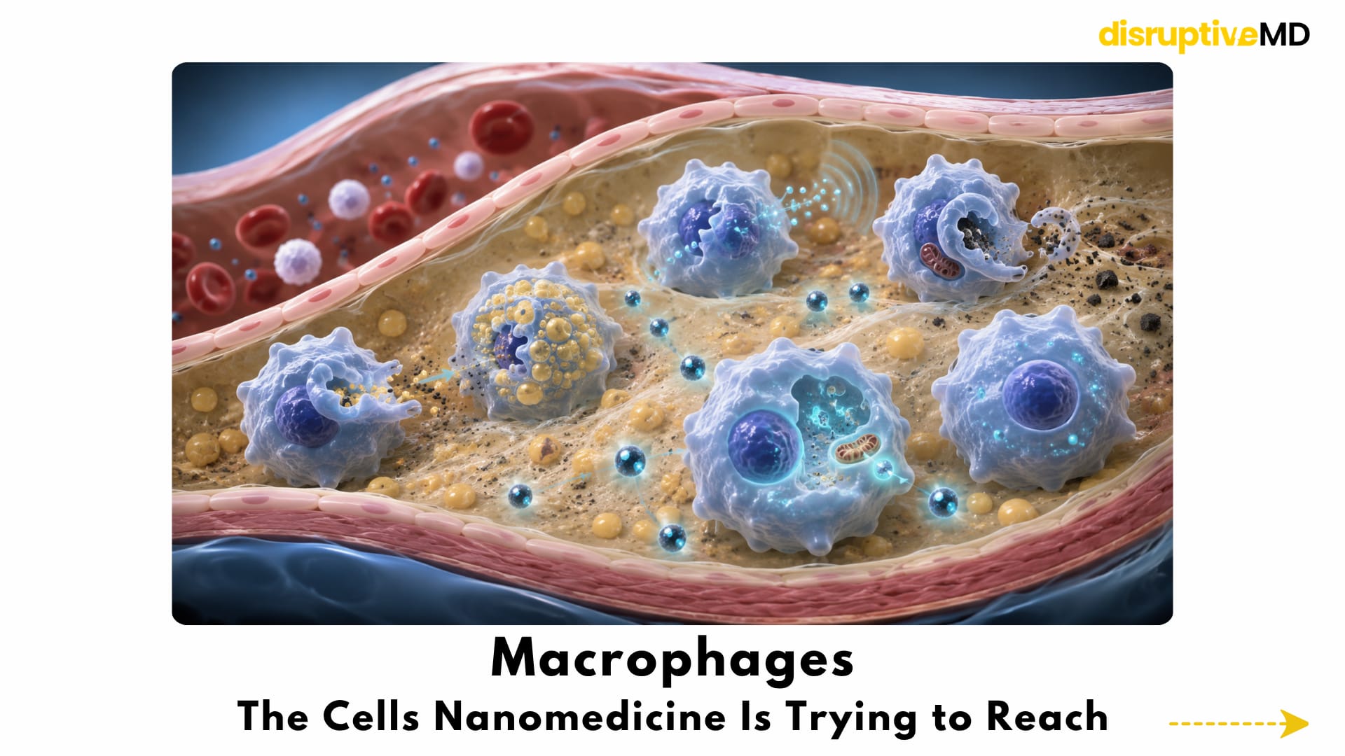
Macrophages: The Cells Nanomedicine Is Trying to Reach
Macrophages sit at the centre of many experimental plaque-targeting strategies.
They help drive the development of atherosclerosis, but they are also essential for resolving inflammation and clearing cellular debris.
This creates an important biological tension.
Macrophages can:
- consume modified cholesterol;
- become lipid-filled foam cells;
- release inflammatory molecules;
- produce enzymes that weaken plaque structure;
- engulf nanoparticles circulating through diseased tissue;
- and, under healthier conditions, clear dead cells and support tissue repair.
Their natural tendency to consume foreign particles makes them appealing targets for nanomedicine.
Researchers are trying to use that behaviour like a biological entry point.
Instead of sending a drug equally throughout the entire body, a particle may be designed so that plaque macrophages preferentially absorb it. Once inside, its payload may influence inflammation, cholesterol handling, oxidative stress, cell survival or tissue cleanup.
Several experimental nanoparticle systems have already reached plaque-associated macrophages in animal models. A small human study also showed that intravenously administered liposomal particles could accumulate within macrophage-rich atherosclerotic tissue, although this did not establish improved cardiovascular outcomes.
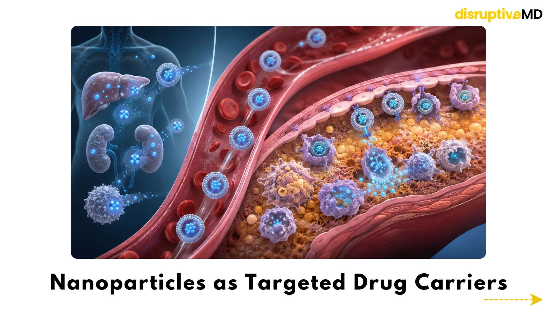
Nanoparticles as Targeted Drug Carriers
Many potentially useful anti-inflammatory or metabolic therapies affect pathways that operate throughout the body.
That creates a major problem.
A drug strong enough to change plaque biology may also affect the immune system, liver, kidneys, blood cells or other tissues.
Nanoparticles may help change that balance.
A therapeutic molecule can be enclosed inside a carrier and protected while it circulates. The surface of the particle can then be modified to improve its interaction with inflamed endothelium, plaque components or immune cells.
Possible payloads include:
- anti-inflammatory drugs;
- lipid-modifying agents;
- inflammation-resolving molecules;
- antioxidants;
- gene-silencing material;
- immune-regulating signals;
- and imaging compounds.
The objective is not merely to make a drug smaller.
It is to control where the drug travels, which cells receive it, when it is released and how long it remains active.
A 2024 preclinical study used antioxidative nanosheets to deliver an inflammation-resolving lipid mediator to macrophages within atherosclerotic lesions. In animal models, the combined system reduced oxidative stress and inflammatory activity while improving features associated with plaque stability. This remains an experimental platform rather than an established patient treatment.
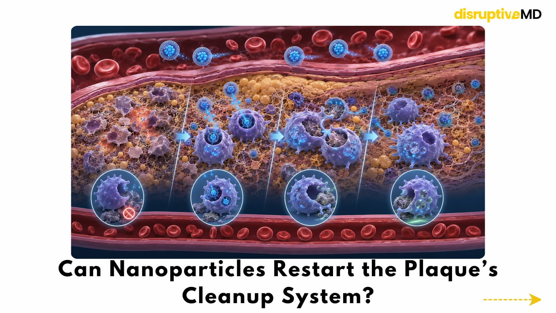
Can Nanoparticles Restart the Plaque’s Cleanup System?
One of the most cutting-edge approaches does not attempt to dissolve the entire plaque.
Instead, it tries to repair the plaque’s failed waste-disposal system.
The process through which macrophages recognise, engulf and remove dead or dying cells is called efferocytosis.
Efficient efferocytosis prevents cellular debris from accumulating and helps inflammation resolve. In advanced atherosclerosis, however, dead cells may display molecular signals that make them harder to engulf.
The result is a plaque filled with material that should have been cleared.
Researchers have therefore developed nanoparticles that enter plaque macrophages and interfere with signals preventing cellular removal.
In a 2020 mouse study, pro-efferocytic nanoparticles were preferentially taken up by lesional macrophages. The treatment improved the clearance of diseased cells and reduced atherosclerotic burden without relying on unrestricted systemic blockade of the same pathway.
A 2024 study moved a related strategy into a porcine model of early atherosclerosis. The nanotherapy reduced the accumulation of apoptotic cells and lowered vascular inflammation without producing the anaemia associated with non-targeted interference in the pathway. This was an important translational step because a large-animal model is biologically closer to human treatment than a mouse model—but it was still not a human outcomes trial.
The idea is powerful because it reframes plaque removal.
The particle is not mechanically vacuuming the artery.
It is helping immune cells resume a cleanup function they have lost.
HDL-Mimetic Nanoparticles: A Biological Cholesterol Shuttle
High-density lipoprotein is associated with reverse cholesterol transport—the movement of excess cholesterol away from peripheral tissues.
This biological role has inspired the development of HDL-mimetic nanoparticles.
These particles attempt to reproduce selected structural or functional properties of natural HDL. Depending on their design, they may interact with plaque macrophages, accept cholesterol or carry therapeutic drugs into inflamed lesions.
Experimental HDL-like systems have been used to deliver statin molecules and other treatments directly toward plaque-associated macrophages. One HDL-mimetic polymeric nanoparticle was designed specifically to target macrophages within atherosclerotic plaques.
Other systems have been designed to respond to enzymes within diseased tissue and release their contents more selectively.
The idea is sometimes described as pulling cholesterol out of the plaque.
But the reality is more complex.
Atherosclerotic plaque contains extracellular lipids, dead cells, fibrous tissue, calcium and altered vascular cells. Removing some cholesterol from macrophages does not instantly erase the entire structure.
HDL-mimetic nanotechnology is better understood as a way of improving cholesterol movement, reducing foam-cell stress and potentially creating a less inflammatory plaque environment.
It is a biological transport strategy, and not a microscopic plaque vacuum.
The Goal May Be Plaque Stability, Not Plaque Disappearance
A treatment does not necessarily need to make a plaque vanish to reduce risk.
A dangerous plaque may become safer if it develops:
- less inflammatory activity
- fewer dying cells
- a smaller necrotic core
- improved cholesterol handling
- more efficient debris clearance
- and a stronger protective fibrous structure
This is the concept of plaque stabilisation.
In a 2020 study, nanoparticles delivered small interfering RNA to macrophages inside mouse plaques. The therapy silenced a molecular pathway associated with defective efferocytosis and improved features of advanced plaque stability.
More recently, multifunctional platforms have combined oxidative-stress control, lipid regulation, immune modulation and targeted drug delivery.
The aim is increasingly not to attack one isolated pathway, but to reshape the plaque environment.
That may prove more realistic than attempting to dissolve decades of arterial disease through a single mechanism.
Smart Nanoparticles That Respond to the Plaque
A plaque has its own microenvironment.
Inflamed lesions may contain increased oxidative stress, altered enzyme activity, acidic regions, dysfunctional immune cells and damaged tissue.
Smart nanoparticles are designed to use those differences as activation signals.
A particle may remain relatively stable during circulation but release its drug more rapidly when it encounters:
- reactive oxygen species;
- specific enzymes;
- altered acidity;
- inflammatory molecules;
- or particular cellular receptors.
This could reduce premature drug release and concentrate treatment within diseased tissue.
A 2026 preclinical system used several levels of targeting. Its outer biological coating helped the particle interact with inflamed arterial tissue, while an additional targeting component directed it toward foam cells. Oxidative stress within those cells then promoted drug release.
In mice, the platform increased cholesterol efflux, reduced inflammatory activity, improved foam-cell clearance and produced features associated with greater plaque stability. It also carried components for multiple imaging methods.
The engineering is impressive.
But every added function also increases manufacturing complexity, testing requirements and the number of ways the platform could behave differently in humans.
A particle that performs perfectly in a controlled mouse model may circulate, degrade or distribute very differently in a person with diabetes, kidney disease, multiple medications and calcified coronary plaques.
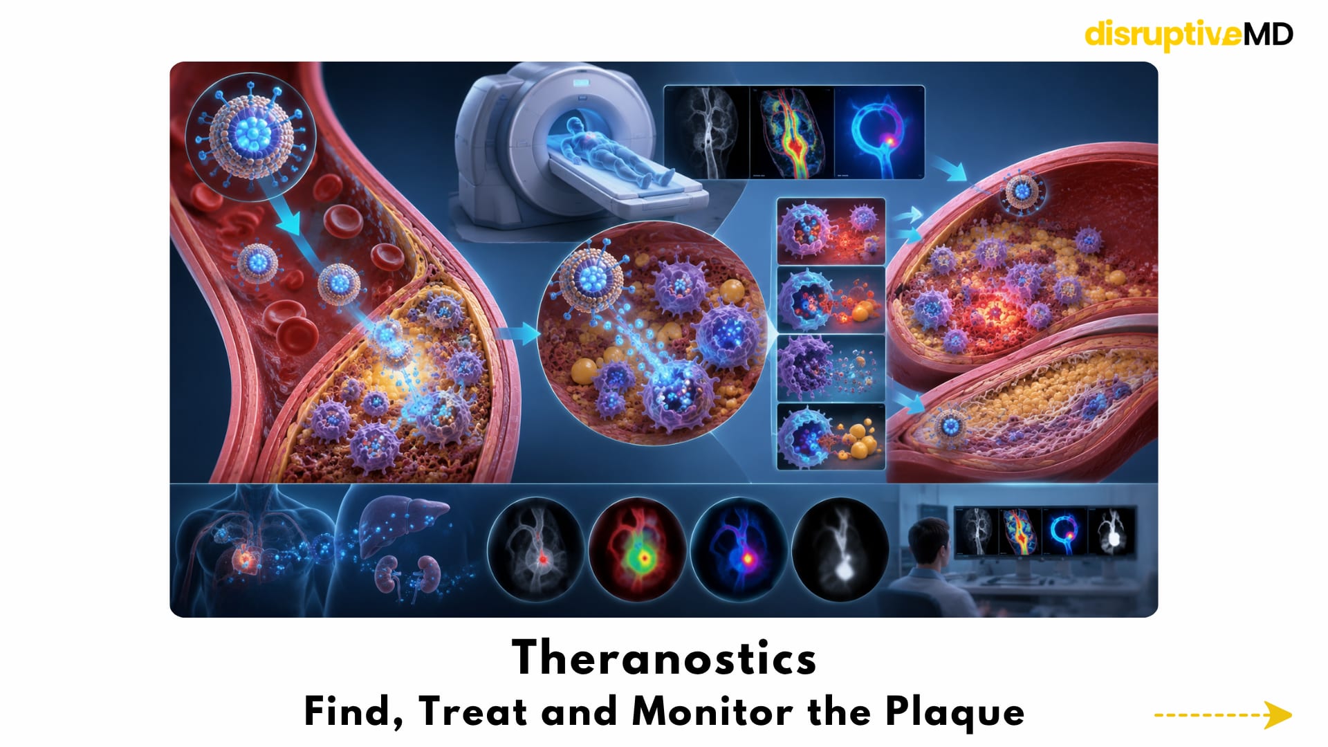
Theranostics: Find, Treat and Monitor the Plaque
Theranostics combines therapy and diagnostics within the same platform.
For atherosclerosis, a theranostic nanoparticle could theoretically:
- travel toward an inflamed plaque;
- create a detectable imaging signal;
- deliver treatment to the lesion;
- and allow the biological response to be monitored.
This could shift arterial imaging away from simply measuring narrowing.
Instead, imaging might reveal:
- macrophage activity
- oxidative stress
- cellular death
- inflammation
- new microvessel formation
- lipid-rich regions
- or changes produced by treatment
Nanoparticle imaging of plaque macrophages has already been demonstrated experimentally, including multimodal PET-CT detection in preclinical atherosclerosis.
The 2026 foam-cell-targeting system took the concept further by integrating magnetic resonance, fluorescence and photoacoustic signals with responsive therapy in mice.
The futuristic workflow would be:
Find the biologically dangerous plaque. Treat its dominant pathway. Image whether the pathway changed.
The difficulty is proving that an imaging signal genuinely predicts a future heart attack or stroke, and that changing that signal improves patient outcomes.
A beautiful scan is not automatically a life-saving treatment.
RNA and Gene-Silencing Delivery
Some of the most important drivers of atherosclerosis operate inside cells.
Traditional drugs may struggle to reach these intracellular targets selectively.
Nanoparticles can carry RNA molecules that reduce the production of specific proteins. This process is often called gene silencing.
In theory, RNA-loaded particles could reduce inflammatory signalling, improve cholesterol export, restore macrophage cleanup or alter other pathways associated with plaque instability.
The 2020 macrophage-targeted siRNA study demonstrated that gene silencing could be delivered to lesional macrophages and improve plaque characteristics in mice.
This is an important proof of concept.
It does not mean plaque-targeted RNA treatment is ready for routine care.
Long-term safety must account for:
- unintended gene silencing
- immune reactions
- distribution outside the plaque
- persistence of the carrier
- liver and spleen accumulation
- dose control
- and the consequences of repeatedly altering immune-cell behaviour
Because atherosclerosis is a chronic disease, a successful therapy may need to remain safe across repeated doses or produce a durable effect without causing long-term immune disruption.
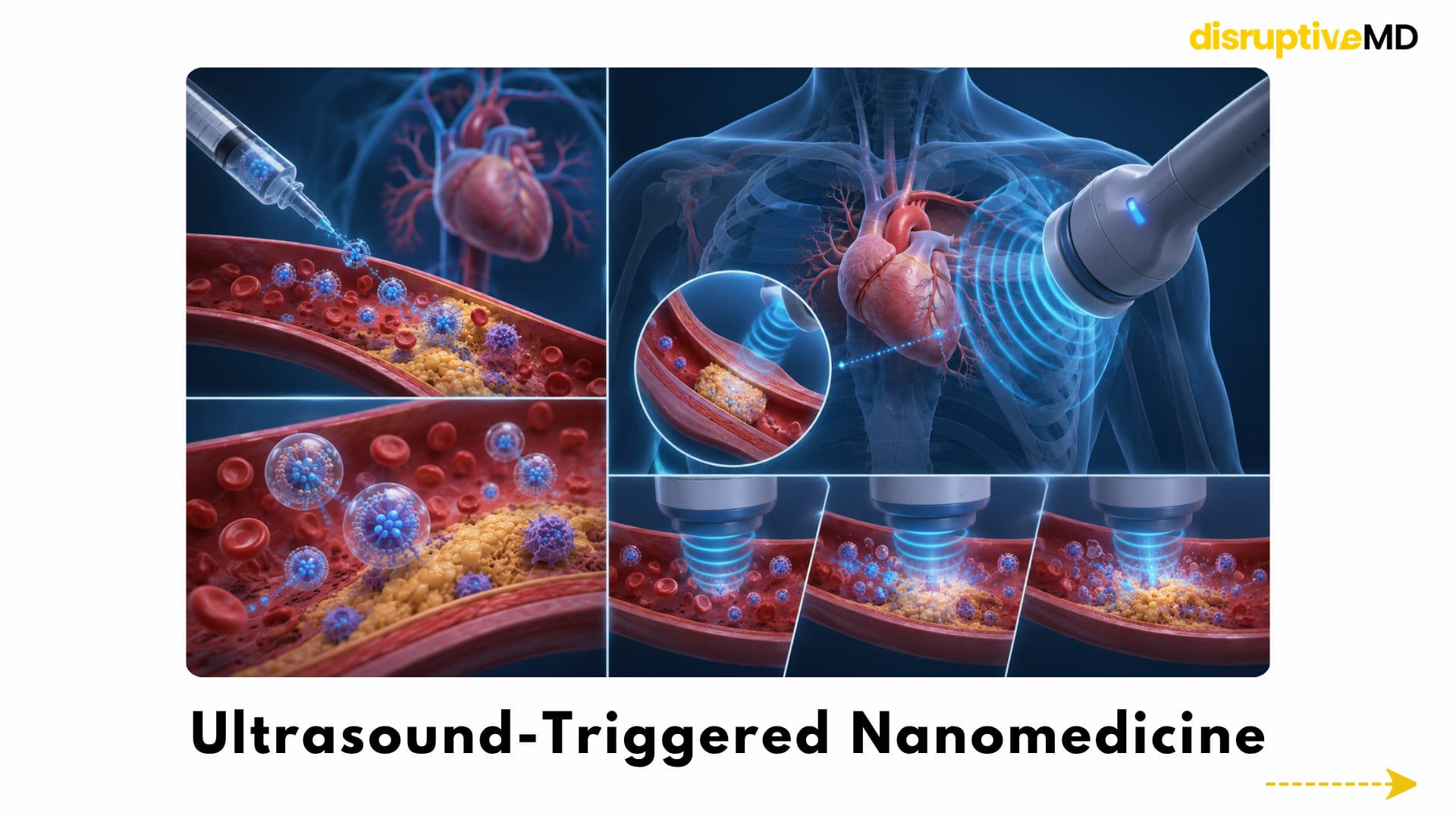
Ultrasound-Triggered Nanomedicine
Another futuristic direction combines nanoparticles with externally applied energy.
Ultrasound could theoretically be used to:
- disrupt drug-loaded microbubbles
- improve movement of a treatment into diseased tissue
- trigger local drug release
- activate a therapeutic compound
- or guide treatment toward a selected arterial region
This creates a possible two-stage system.
First, the nanomedicine is injected.
Then ultrasound is focused near the target plaque to increase or activate treatment locally.
The concept could offer greater spatial control than an injection alone.
However, coronary arteries move with the beating heart, plaques may sit deep within the chest, and ultrasound energy must be delivered without damaging the vessel, blood cells or surrounding tissue.
Ultrasound-triggered plaque nanotherapy therefore remains an experimental direction rather than a validated method for cleaning coronary arteries.
When Targeting Becomes Too Complicated
Theoretically, a nanoparticle can be designed to recognise almost any biological feature.
In practice, the bloodstream is an extremely difficult environment.
Proteins rapidly attach to nanoparticle surfaces, potentially changing how the particle behaves. The liver, spleen and immune system may remove it before it reaches the plaque.
The diseased endothelium may not allow uniform entry.
One plaque may be dominated by macrophage inflammation, while another is heavily calcified and fibrotic. A treatment designed for one plaque phenotype may have little effect on another.
Other challenges include:
- nanoparticle aggregation
- complement or immune activation
- toxicity of degradation products
- accumulation in non-target organs
- inconsistent manufacturing
- limited penetration into advanced plaque
- differences between animal and human arteries
- and uncertainty about repeated dosing
Researchers must also prove that the treatment does more than improve microscopic plaque features.
A clinically meaningful therapy must ultimately reduce outcomes such as heart attack, stroke, urgent procedures or cardiovascular death without introducing unacceptable harm.
That is a much higher standard than shrinking a plaque in a mouse.
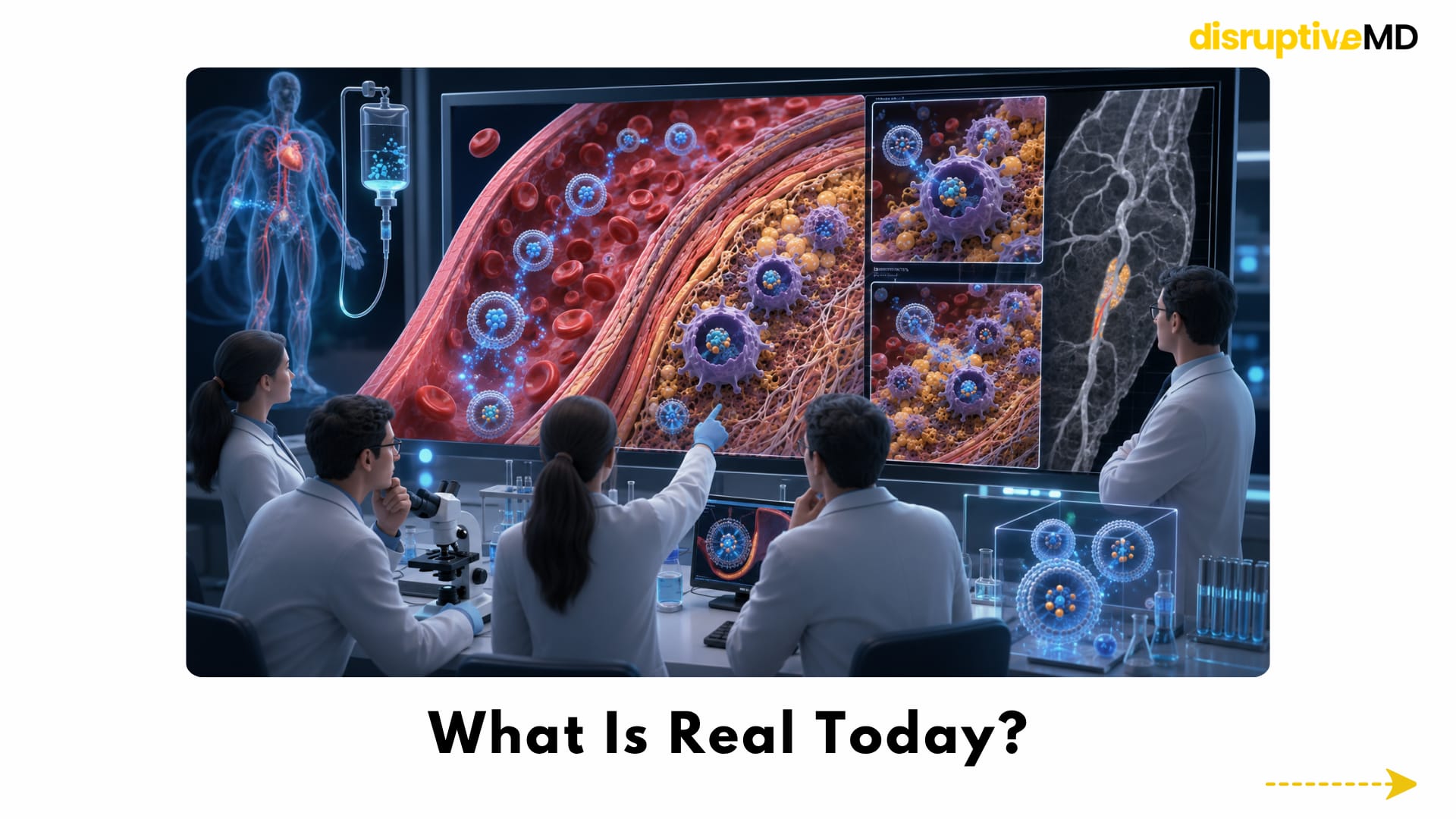
What Is Real Today?
Nanomedicine for atherosclerosis is a legitimate and active research field.
What is currently real includes:
- nanoparticles engineered to carry therapeutic and imaging payloads
- preferential uptake of certain particles by plaque macrophages
- experimental delivery of anti-inflammatory drugs and inflammation-resolving molecules
- nanoparticle-based RNA silencing in plaque cells
- HDL-like particles designed to influence cholesterol transport
- responsive particles activated by aspects of the plaque microenvironment
- nanoparticle imaging of macrophage-rich lesions
- promising results in cell, mouse and other animal models
- and limited human studies demonstrating that selected nanocarriers can reach atherosclerotic tissue
The 2024 porcine efferocytosis study is particularly important because it tested targeted macrophage therapy in a large-animal model and reported reduced lesion inflammation without anaemia.
A human study published in 2015 demonstrated plaque accumulation of intravenously administered liposomal prednisolone, showing that systemic nanocarriers can reach human atherosclerotic macrophages. It did not prove that this strategy prevents heart attacks or produces meaningful plaque regression.
What Is Not Fully Real Yet?
There is currently no routine infusion that safely locates every dangerous plaque and cleans it from the inside.
The following remain unproven or misleading:
- nanobots scraping cholesterol from artery walls
- a nanoparticle vacuum that removes advanced plaque in one treatment
- routine plaque-targeted RNA therapy
- a clinically validated smart particle that activates only inside coronary plaques
- a proven theranostic infusion that finds, treats and monitors vulnerable plaques
- replacement of established cholesterol-lowering and risk-reduction treatment
- replacement of stents or bypass surgery when mechanical restoration of blood flow is required
- and demonstrated prevention of heart attacks or strokes through an advanced plaque-targeted nanotherapy
Most sophisticated systems remain preclinical.
Some have been studied in large animals. A small number of simpler nanocarrier or nanoparticle-imaging concepts have reached human research.
That is meaningful progress, but it is not the same as everyday clinical treatment.
Fact Base
Targeted efferocytosis has strong preclinical support
Nanoparticles have been used to enter plaque macrophages, suppress signals that prevent cellular engulfment and improve removal of dead plaque cells in mice. A related formulation later reduced lesion inflammation and apoptotic-cell accumulation in a porcine model without causing anaemia.
Macrophage-targeted RNA delivery can alter plaque biology
Small interfering RNA delivered through nanoparticles improved efferocytosis and features of plaque stability in mouse atherosclerosis.
Inflammation-resolving molecules can be carried into plaques
Antioxidative nanosheets carrying an inflammation-resolving lipid mediator reduced oxidative stress and inflammatory plaque features in preclinical models.
HDL-inspired particles can target plaque macrophages
Synthetic HDL-like particles have been engineered to interact with plaque macrophages and carry therapeutic compounds. These remain experimental and should not be interpreted as devices that physically suction plaque from the vessel wall.
Theranostic systems can combine imaging and treatment
A 2026 mouse study combined multimodal plaque imaging, foam-cell targeting, oxidative-stress-responsive drug release, cholesterol efflux and improved efferocytosis within one nanoparticle platform.
Human targeting has been demonstrated, but clinical benefit has not
Intravenous liposomal particles have been shown to accumulate in human plaque macrophages. This established biological delivery—not prevention of cardiovascular events.
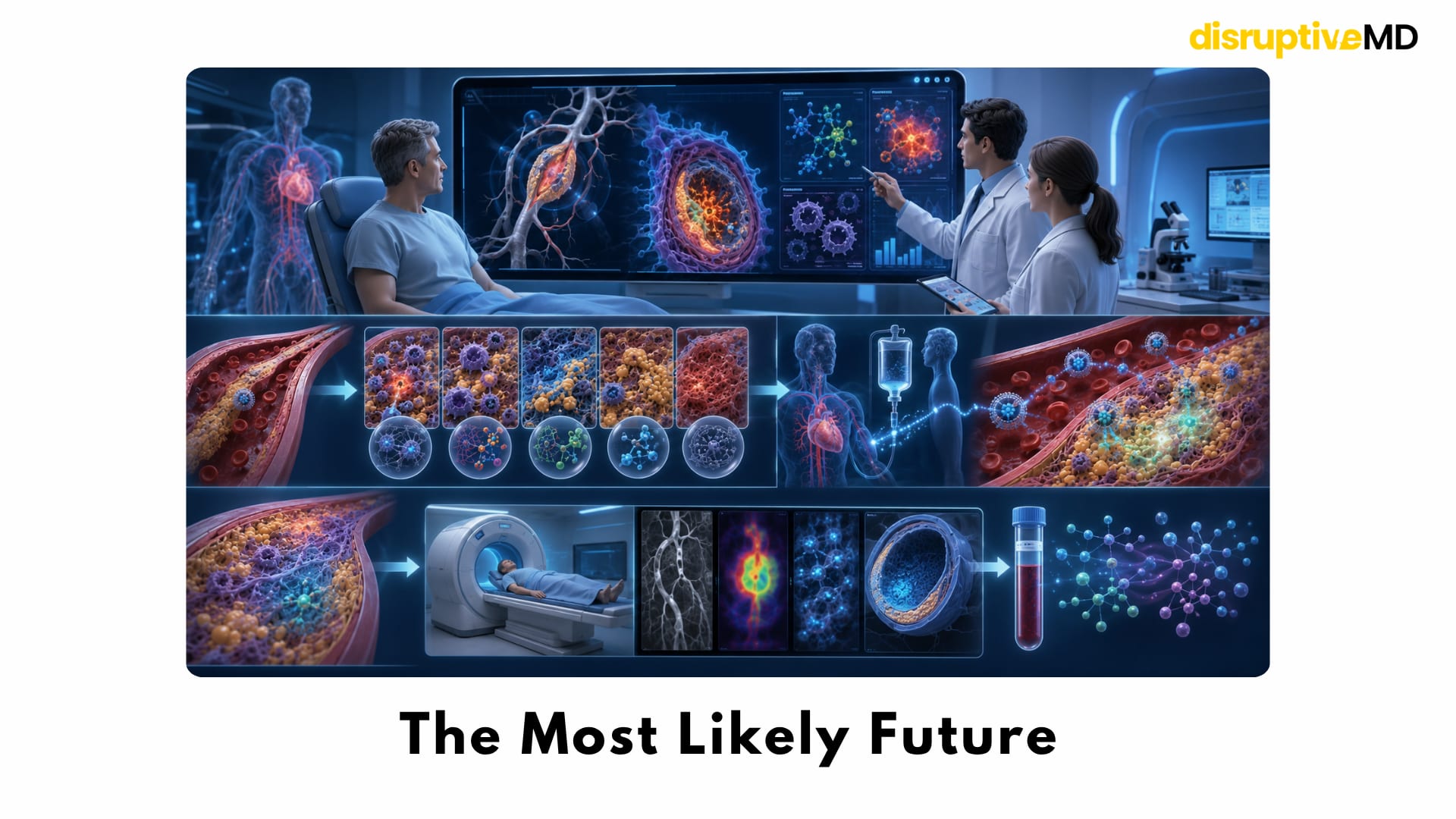
The Most Likely Future
The first successful plaque nanotherapies may not be universal artery-cleaning treatments.
They may be used in selected patients with a particular biological plaque profile.
A future workflow could involve advanced imaging or blood-based risk markers identifying persistent arterial inflammation despite standard treatment.
A nanotherapy could then be selected according to the dominant problem:
- excessive macrophage inflammation
- impaired efferocytosis
- defective cholesterol export
- high oxidative stress
- or another molecular feature
The treatment might be administered for a limited period and its response assessed through molecular imaging or circulating biomarkers.
That is more plausible than releasing autonomous machines into the bloodstream and asking them to scrape away every plaque.
The future is likely to be pharmacological, biological and image-guided, and not mechanical science fiction.
Key Takeaway
Nanotechnology is unlikely to clean arteries in the way a brush cleans a pipe.
Its true potential is more sophisticated.
It may help treatments travel directly to diseased plaque, calm harmful inflammation, remove cholesterol from overloaded cells, restart the clearance of dead tissue, strengthen vulnerable lesions and reveal whether a plaque is biologically responding.
The greatest breakthrough may not be making every plaque disappear.
It may be preventing the dangerous plaque from rupturing.
The real future of nanotechnology in arteries is not teaching machines to scrape the vessel wall - it is teaching the artery wall to heal before plaque becomes deadly.
References
- Flores AM, et al. Pro-efferocytic nanoparticles are specifically taken up by lesional macrophages and prevent atherosclerosis. 2020. DOI: 10.1038/s41565-019-0619-3.
- Bamezai S, et al. Pro-efferocytic nanotherapies reduce vascular inflammation without inducing anemia in a large animal model of atherosclerosis. 2024. DOI: 10.1038/s41467-024-52005-1.
- Tao W, et al. siRNA nanoparticles targeting CaMKIIγ in lesional macrophages improve atherosclerotic plaque stability in mice. 2020. DOI: 10.1126/scitranslmed.aay1063.
- He Z, et al. Resolvin D1 delivery to lesional macrophages using antioxidative black phosphorus nanosheets for atherosclerosis treatment. 2024. DOI: 10.1038/s41565-024-01687-1.
- Song J, et al. A hierarchical theranostic nanoagent for multimodal imaging and targeted foam cell intervention in atherosclerosis. 2026. DOI: 10.1038/s41467-026-70463-7.
- Li Y, et al. A programmable platelet theranostic platform for adaptive multi-stage delivery and synergistic immunotherapy in atherosclerosis. 2025. DOI: 10.1038/s41467-025-61789-9.
- van der Valk FM, et al. Prednisolone-containing liposomes accumulate in human atherosclerotic macrophages upon intravenous administration. 2015. DOI: 10.1016/j.nano.2015.02.021.
- Sanchez-Gaytan BL, et al. HDL-mimetic PLGA nanoparticle to target atherosclerosis plaque macrophages. 2015. DOI: 10.1021/bc500517k.