
Introduction
In vitro fertilization (IVF) has for years been one of the most remarkable procedures in medicine: mix eggs and sperm outside the body, grow embryos in a lab, and pick one embryo to transfer to the uterus. For decades, many of the most significant decisions in IVF have hinged on the judgment of experts: how an egg looks, how a sperm swims, how an embryo divides, and which embryo looks most likely to lead to pregnancy. IVF 2.0 is changing all that. The new generation of reproductive medicine is integrating artificial intelligence, robots, non-invasive testing, stem-cell technology, and better fertility preservation into a discipline that was nearly totally manual.
Technology is taking the place of fertility specialists or embryologists. The more plausible future is that IVF becomes more quantifiable, standardized, data-driven, and physiologically ambitious. A few of these developments are already serving as decision assistance tools. Some are early technology for the clinic. Others are experimental and should not be promoted as a proven treatment. The actual revolution is that IVF is going from just “creating embryos in a lab” to understanding, selecting, maintaining, and potentially altering reproductive cells with far better precision.
- IVF is shifting from manual judgment toward measurable reproductive intelligence.
- AI, robots, testing, and preservation are changing lab decision-making.
- Technology supports specialists, but does not replace clinical responsibility.
1. AI-powered embryo selection
AI-powered embryo selection employs computer algorithms to analyse embryo photos and determine which embryo may have the best likelihood of implantation, pregnancy, or live birth. In traditional IVF, embryologists analyse embryos based on morphology (visible structure of the embryo): quantity and arrangement of cells, appearance of the inner cell mass, trophectoderm, blastocyst expansion, fragmentation, and timing of development. This expert visual grading is still crucial, but it can be subjective, as various embryologists may read the same embryo somewhat differently. AI seeks to decrease that subjectivity by translating embryo appearance and time-lapse development into quantitative patterns.
In practice, AI systems are typically trained on vast sets of photos of embryos, connected to known outcomes. The software would be able to assess static images from microscopes, time-lapse films from incubators, blastocyst morphology, timing of development, or aspects that are too subtle for human eyes to quantify consistently. Some recent models also attempt to estimate the likelihood that an oocyte will subsequently develop into a euploid blastocyst, i.e., one with the predicted number of chromosomes. In 2025, a Human Reproduction abstract described a multicenter external validation of an AI model that used images of mature oocytes, age, and associated features to estimate the probability of euploid blastocyst development, but the authors also stated that the work was retrospective and needed prospective evaluation before routine clinical use.
AI embryo selection should be considered an adjunct of clinical practice, not a decision-maker. Some clinics utilize embryo-scoring software to assist embryologists, increase uniformity, and reduce the time used to examine and help rank embryos when there is more than one good-quality embryo. It may be particularly beneficial in combination with time-lapse incubation, since embryos can be seen without repeatedly removing them from the incubator. But AI is not a guarantee of conception, is not a replacement for genetic testing, and cannot take into account uterine factors, sperm factors, maternal health, lab conditions, or chance. The evidence is encouraging but mixed.* In a large 2024 randomized, double-blind, non-inferiority trial in Nature Medicine, deep learning embryo selection was compared with traditional morphology-based selection across 14 IVF facilities. Pregnancy and live birth outcomes were largely similar; however, the study did not demonstrate non-inferiority of the AI approach to standard morphology by its prespecified parameters. It did demonstrate a consistent, user-independent technique and a considerable reduction in assessment time, which may be of importance for laboratory workflow.*
The good news of AI is IVF might go from "Which embryo looks best?" Which embryo has the best data profile?” The catch is that many AI algorithms are trained on curated datasets and may not perform equally well across different clinics, populations, incubation systems, picture kinds, or laboratory techniques. AI models can also be difficult to explain, raising ethical and medico-legal issues if patients are told that a “score” impacted the choice of embryo.
AI embryo selection is not creating artificial life. It is pattern recognition, applied to the examination of embryos. The future hinges on prospective studies, transparency, bias testing, and evidence of improvement in relevant outcomes like live birth, and not simply prediction scores.
2. Robotic and automated IVF laboratories
IVF labs with robotics and automation are designed to eliminate the manual unpredictability of IVF lab work. IVF relies on very delicate procedures: identifying oocytes, preparing sperm, selecting sperm, inserting a sperm into an egg (ICSI), transporting embryos across media, storing embryos or eggs, thawing them, and maintaining precise environmental conditions. These steps are completed by highly trained embryologists, yet even in the hands of experts, IVF is technically hard and labour consuming. Automation tries to make these procedures more reproducible.
Robotic IVF systems use computer vision, microfluidics, robotic arms, digital micromanipulators, lasers, and AI-directed control systems. The system can recognize structures, position instruments, manage the depth of injection, stabilize sperm or oocytes, and standardize repetitious operations, rather than an embryologist manually manipulating each tool under a microscope. A peer-reviewed case report in Reproductive Biomedicine Online in 2025 described a digitally controlled, remotely operated ICSI device that automated the micromanipulation portion of ICSI and resulted in a live delivery. The system still needed human setup and supervision, and only part of the process was autonomous, but it showed practicality.
The technology is not a global standard for everyday use but is utilized largely for research, early clinical investigations, and some experimental applications. Some firms are developing integrated, automated IVF platforms that promise to perform numerous steps from egg and sperm preparation to fertilization and cryopreservation. For example, Conceivable Life Sciences says its AURA platform is an AI-powered automated IVF facility that can complete all the stages required to unite egg and sperm and make an embryo. Robotic IVF experiments and early births have also been reported independently; however, these technologies are still being evaluated and have not yet convincingly outperformed conventional expert IVF.
The upside might be huge: more standardization of IVF facilities, less reliance on the unique skills of operators, less fatigue, better documentation, remote expert supervision, and wider access in areas with limited embryology knowledge. Automation can also increase quality control. Every movement, timing decision, and environmental change can be recorded. This could, eventually, transform IVF from an artisanal manual operation into a regulated biological manufacturing process.
Risks and constraints are just as crucial. Robotic systems can fail, need servicing, bring new technical faults, and can be expensive to set up. A robot doing a delicate step “successfully” once does not mean it improves outcomes for hundreds of patients. Fertility treatment also involves emotions, ethics, counselling, consent, and human decision-making that cannot be automated.
Robotic IVF is not a robot creating babies on its own. It is the automation of some steps in the lab under medical, embryology, and regulatory supervision. The disruptive aspect is not sci-fi but standardization.
3. Non-invasive embryo testing
Biopsy is the typical method used for standard preimplantation genetic testing, or PGT. In PGT-A, a few cells are taken from the trophectoderm, the outer layer of a blastocyst that eventually forms part of the placenta, and those cells are examined for chromosomal abnormalities. The biopsy-based technique has been widely employed in selected IVF cases but involves a certain level of technical expertise, specialized equipment, manipulation of the embryo, and cautious counselling.
Non-invasive embryo testing attempts to collect genetic information without taking cells away from the embryo. The major way is to examine the cell-free DNA produced by the embryo in the wasted culture medium, the fluid in which the embryo has been growing. In theory, the embryo drips a few bits of DNA that can be collected and sequenced.
In research practice, embryologists collect discarded culture material at a certain time, commonly around the blastocyst stage, and then the DNA is amplified and examined using genetic testing tools. Some methods additionally contain washing processes of embryos to reduce contamination. Results can be affected by time, culture conditions, washing procedure, fertilization method, DNA quantity, and contamination. A 2024 study indicated that day-6 collection and sequential washing increased concordance with trophectoderm biopsy, but the authors said more randomized trials are needed before non-invasive PGT may be used as a regular embryo selection technique.
The appeal is clear: genetic information without a biopsy. This may lessen technical burden, avoid manipulation of the embryo, and make testing more accessible. It could also be a beneficial adjunctive tool when biopsy is not possible or when clinics desire extra non-invasive information. The disruptive viewpoint is that in the future, embryo assessment may include morphology, AI imaging, metabolic signals, and cell-free DNA rather than relying on biopsy.
But the evidence is full of big holes. In 2025, Scientific Reports said non-invasive PGT-A is biologically attractive but is still limited in terms of maternal DNA contamination, fragmented DNA, unpredictable DNA shedding, imprecise representation of the true chromosomal condition of the embryo, and inconsistent diagnostic performance. The study also pointed out that skipping biopsy does not necessarily mean better safety or better IVF outcomes.
Currently, non-invasive embryo testing should be considered as an emerging technique, rather than a complete replacement for verified biopsy-based PGT. The greatest risk is that of misclassification: that is, a normal embryo might be improperly classified as abnormal, or an aberrant embryo might be wrongly classified as suitable. Students should learn that “non-invasive” does not equal “better.” In reproductive medicine, a less intrusive test must still be reliable, therapeutically helpful, reproducible, and ethically safe.
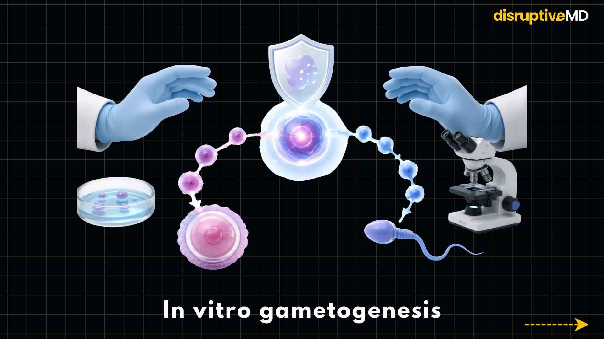
4. In vitro gametogenesis
In vitro gametogenesis, or IVG, is an effort to produce eggs or sperm from stem cells in the lab. This is one of the most groundbreaking concepts in the field of reproductive medicine, as it goes beyond collecting eggs and sperm from the body. Instead, scientists are investigating whether ordinary cells may be re-programmed to become induced pluripotent stem cells and then be directed to produce functional gametes.
The basic idea starts with a somatic cell, such as a skin or blood cell. They revert it to something like a stem cell and then try to guide it through the complex biological processes that normally occur in the ovaries or testes. This indicates that for sperm, this results in cells competent to undergo meiosis, proper chromosomal reduction, imprinting, and conception. The task is significantly more challenging for eggs, as the egg is a big, highly specialized cell with unique cytoplasm, mitochondria, epigenetic programming, and development capability. IVG is not merely “making a cell that looks like an egg or sperm.” It is developing a gamete that can contribute half the genome safely and support healthy embryo development.
IVG is not the conventional IVF in clinical practice today. It is not an authorized fertility therapy for use in human reproduction. Future potential applications are age-associated infertility, cancer-related infertility, premature ovarian failure, lack of gametes, hereditary infertility, and the prospect of genetically related children for same-sex couples. But those are still future possibilities, not current clinical options. A recent study in Human Reproduction in 2025 identified stem-cell-derived gametes as a nascent technology and highlighted the need to address safety, efficacy, ethics, legality, accountability, and long-term monitoring before clinical implementation.
Recent lab work explains why the field is intriguing but also why it needs to be approached with caution. In 2025, Oregon Health & Science University published proof-of-concept research that involved inserting human skin-cell nuclei into donor oocytes and prompting them to jettison half their chromosomes before conception. While some embryos reached the blastocyst stage, most were defective or chromosomally challenged, and the researchers noted much more work needs to be done before any therapeutic use. Reuters also noted that major safety issues remain and that researchers predict another decade or more of research before the strategy might be considered for human trials, if clinical trials are allowed.
The potential advantage of IVG is huge: it could extend reproductive options for patients who cannot generate acceptable eggs or sperm. It may also allow scientists to examine early human development, reasons for infertility, meiotic mistakes, and the biology of gametes in ways that are challenging at present. The risks are just as serious: chromosomal aberrations, epigenetic errors, imprinting problems, unknown consequences on kids, exploitation of embryos, unequal access, and difficult ethical issues about parenthood and genetic selection.
IVG is not “advanced IVF” as it is understood in the clinical setting. It’s a potential future platform that could change the meaning of IVF. If safe and regulated, IVF may go beyond collecting gametes to creating gametes. But today, IVG is mostly on the research front.
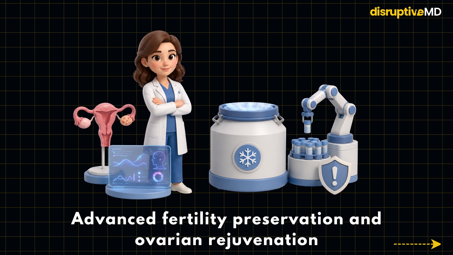
5. Advanced fertility preservation and ovarian rejuvenation
Advanced fertility preservation is an extension of IVF technology from treating infertility to protecting future fertility. Oocyte cryopreservation, or egg freezing, is the most well-established aspect of this field. After ovarian stimulation, eggs are harvested and frozen using a fast-freezing procedure known as vitrification, which minimizes damage from ice crystals. Embryo freezing and ovarian tissue cryopreservation are extremely crucial, particularly for those undergoing cancer therapy, gonadotoxic medications, or illnesses that may impact fertility.
Egg freezing is no longer considered experimental within the appropriate clinical conditions. ASRM points out that oocyte cryopreservation was first considered experimental, but the experimental designation was removed after advances in laboratory methods and comforting outcome data. ESHRE’s guideline on female fertility preservation also mentions fertility preservation options like oocyte, embryo, and ovarian tissue cryopreservation and states that oocyte cryopreservation has now become the preferred method for many women wanting fertility preservation for age-related or medical reasons, thanks to vitrification.
The latest IVF 2.0 layer is the use of AI and automation to make fertility preservation more predictive and consistent. AI-assisted oocyte assessment could allow the egg quality to be evaluated from photographs. Automated cryopreservation methods may mitigate handling variance. Ovarian reserve prediction models may help forecast how someone may respond to stimulation or how many eggs may be recovered. While these techniques do not ensure future pregnancy, they may help clinicians counsel patients in a more realistic manner and personalize treatment.
Established fertility preservation is accompanied by experimental “ovarian rejuvenation” techniques. These include platelet-rich plasma, or PRP; stem-cell-related techniques; mitochondrial treatments; and ovarian tissue activation. PRP entails collecting the platelets rich in growth factors from a patient’s blood and injecting them into the ovarian tissue with the hope of boosting the ovarian function. Reviews discuss putative molecular causes such as better angiogenesis, reduced oxidative stress, and mitochondrial effects but underline the necessity for standardized protocols and controlled trials before PRP can be regarded as a validated fertility treatment.
Mitochondrial and stem-cell approaches are even more experimental. Earlier attempts at mitochondrial transfer did not show a significant benefit for embryo quality in controlled tests, and some have been abandoned. Stem-cell methods for premature ovarian insufficiency and decreased ovarian reserve are still under investigation. Numerous promising preclinical or early clinical signals but not enough proof to be used routinely.
The potential benefit of this whole area is that IVF could form part of fertility lifespan management. Reproductive medicine may increasingly assess risk and preserve eggs or tissue sooner, using predictive technologies to guide timing rather than waiting until infertility is confirmed. The downside is that fertility prediction is still imprecise, egg freezing does not necessarily equal a baby, and “ovarian rejuvenation” can be promoted quicker than it can be scientifically validated. Egg freezing and embryo freezing are recognized tools for fertility preservation, and ovarian rejuvenation is yet exploratory and should be presented carefully.
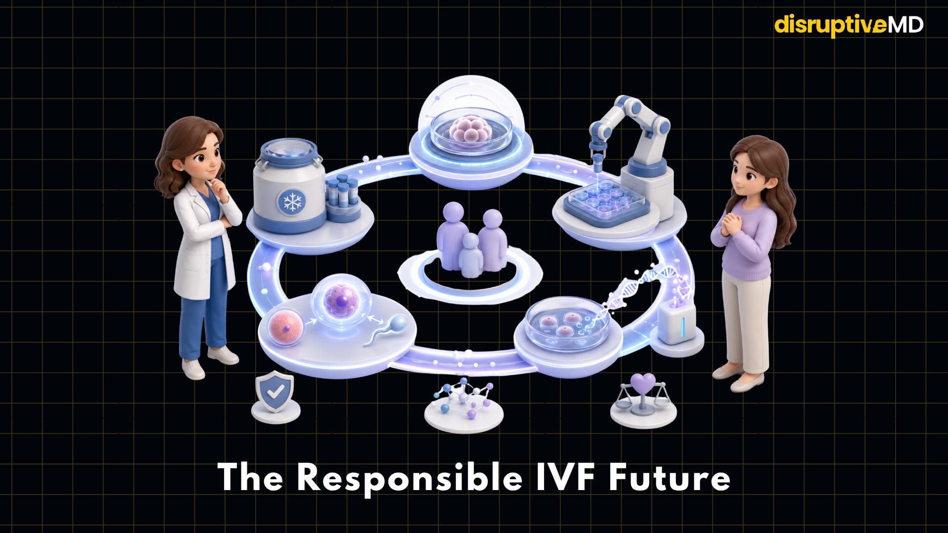
Clinical impact
IVF 2.0 is a move from observation to intelligence measurement in reproductive medicine. AI could aid with more consistent embryo ranking. Robotics could automate sensitive lab tasks. In the future, non-invasive diagnostics may eliminate the need to biopsy embryos. One day, IVG might also challenge the notion that eggs and sperm have to be taken from the body. Advanced fertility preservation could transform IVF from a last step after infertility to a proactive strategy for reproductive planning.
The responsible message, however, is that not every cutting-edge new IVF technology is suitable for routine clinical usage. AI embryo selection is promising but currently being confirmed. Robotic IVF is not yet proven superior at scale. Non-invasive embryo testing is appealing but not yet reliable enough to replace traditional PGT in many scenarios. IVG is still experimental. Although egg freezing is an established procedure, ovarian rejuvenation is yet exploratory.
The future IVF clinic may appear significantly different from today’s IVF clinic. It might start with predicted ovarian modelling, go through AI-assisted oocyte and embryo assessment, involve robotic lab handling, and merge imaging, genetics, and biology into a more personalized treatment approach.
References
- Illingworth, P.J., Venetis, C., Gardner, D.K. et al. Deep learning versus manual morphology-based embryo selection in IVF: a randomized, double-blind noninferiority trial. Nat Med 30, 3114–3120 (2024). https://doi.org/10.1038/s41591-024-03166-5.
- N Mercuri, J Fjeldstad, S Corsac, D Nayot, A Krivoi, O-004 Multi-center, external validation of a novel artificial intelligence (AI) model that predicts blastocyst PGT-A results from mature oocytes, Human Reproduction, Volume 40, Issue Supplement_1, June 2025, deaf097.004, https://doi.org/10.1093/humrep/deaf097.004.
- Mendizabal-Ruiz G, et al. A digitally controlled, remotely operated ICSI system: case report of the first live birth. Reproductive BioMedicine Online, 2025.
- ESHRE. Good practice recommendations for preimplantation genetic testing.
- Chow, J.F.C., Lam, K.K.W., Cheng, H.H.Y. et al. Optimizing non-invasive preimplantation genetic testing: investigating culture conditions, sample collection, and IVF treatment for improved non-invasive PGT-A results. J Assist Reprod Genet 41, 465–472 (2024). https://doi.org/10.1007/s10815-023-03015-3.
- Wu, TF., Lee, CI., Shih, HH. et al. Extended blastocyst culture improves DNA yield in non-invasive preimplantation genetic testing for aneuploidy but diagnostic specificity remains limited. Sci Rep 15, 34906 (2025). https://doi.org/10.1038/s41598-025-18764-7.
- Ilse J de Bruin, Merel M Spaander, Simone Harmsen, et al. on behalf of the HipGametes Consortium, Stem cell-derived gametes: what to expect when expecting their clinical introduction, Human Reproduction, Volume 40, Issue 9, September 2025, Pages 1605–1615, https://doi.org/10.1093/humrep/deaf123.
- ASRM. Planned oocyte cryopreservation and evidence-based outcomes after oocyte cryopreservation.
- The ESHRE Guideline Group on Female Fertility Preservation, Richard A Anderson, Frédéric Amant, Didi Braat, et al. ESHRE guideline: female fertility preservation, Human Reproduction Open, Volume 2020, Issue 4, 2020, hoaa052, https://doi.org/10.1093/hropen/hoaa052.
- Ahmadi F, et al. PRP and ovarian function/oocyte quality review. Journal of Reproductive Immunology, 2025.