
What if a microscope could help detect cancer, grade disease, predict risk, and guide treatment decisions?
Pathology is one of the most consequential parts of modern medicine.
A tiny tissue sample can determine whether cancer is present, identify its type, show how aggressively it is behaving, measure treatment-related biomarkers and influence decisions about surgery, chemotherapy, targeted treatment or immunotherapy.
Traditionally, this information has depended on an expert pathologist examining stained tissue through a microscope. The pathologist studies cell shape, tissue architecture, invasion, inflammation, necrosis, mitotic activity and dozens of other visual clues before combining them with clinical and laboratory information.
Artificial intelligence does not remove that expertise.
It adds a new analytical layer.
Once glass slides are converted into high-resolution digital images, AI can scan enormous areas of tissue, highlight suspicious regions, count cells, measure staining, compare patterns and detect relationships that may be difficult to recognise consistently with the human eye alone.
The goal is not to replace the pathologist.
The real goal is:
To make pathology faster, more consistent, more measurable and more connected to precision medicine.
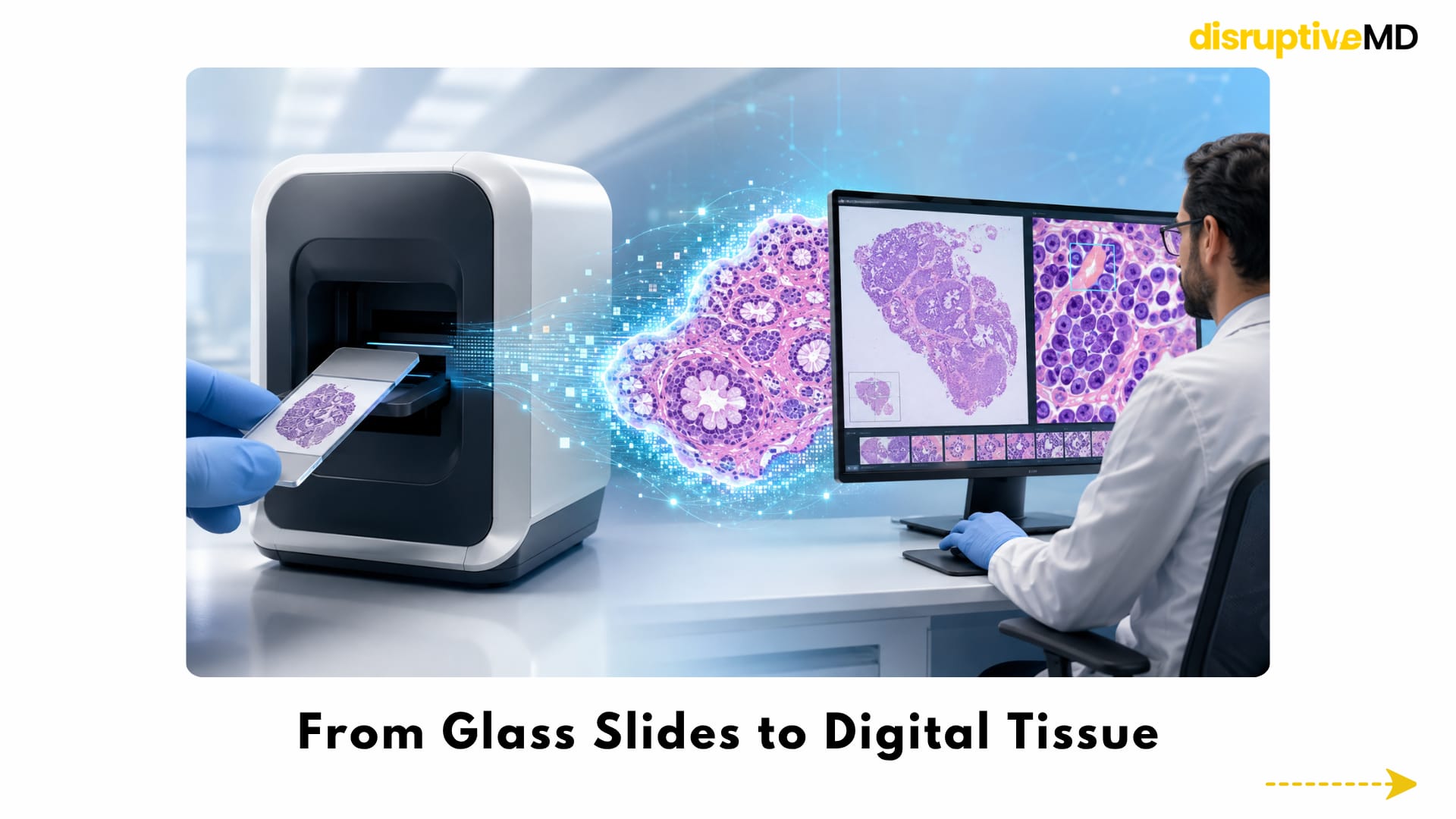
- In digital pathology, a prepared glass slide is scanned to create a whole-slide image.
- The pathologist can move across the image, change magnification and inspect individual cells on a computer screen.
- The glass slide remains the biological source. The digital image turns that source into data.
From Glass Slides to Digital Tissue
The first revolution in AI pathology is not the algorithm.
It is digitisation.
In digital pathology, a prepared glass slide is scanned to create a whole-slide image. This is a high-resolution digital representation of the entire tissue section. The pathologist can move across the image, change magnification and inspect individual cells on a computer screen in much the same way that a conventional microscope is navigated.
Large validation studies have shown that properly validated whole-slide imaging can support primary pathological diagnosis with performance comparable to conventional microscopy.
Digitisation changes what can be done with the slide. The image can be:
- reviewed remotely
- securely shared for specialist opinions
- stored and retrieved electronically
- compared with earlier specimens
- incorporated into teaching and quality-control systems
- analysed computationally
The glass slide remains the biological source. The digital image turns that source into data.
Without digital pathology, AI has very little to analyse. With it, pathology becomes a computational discipline as well as a visual one.
How AI Reads an Entire Pathology Slide
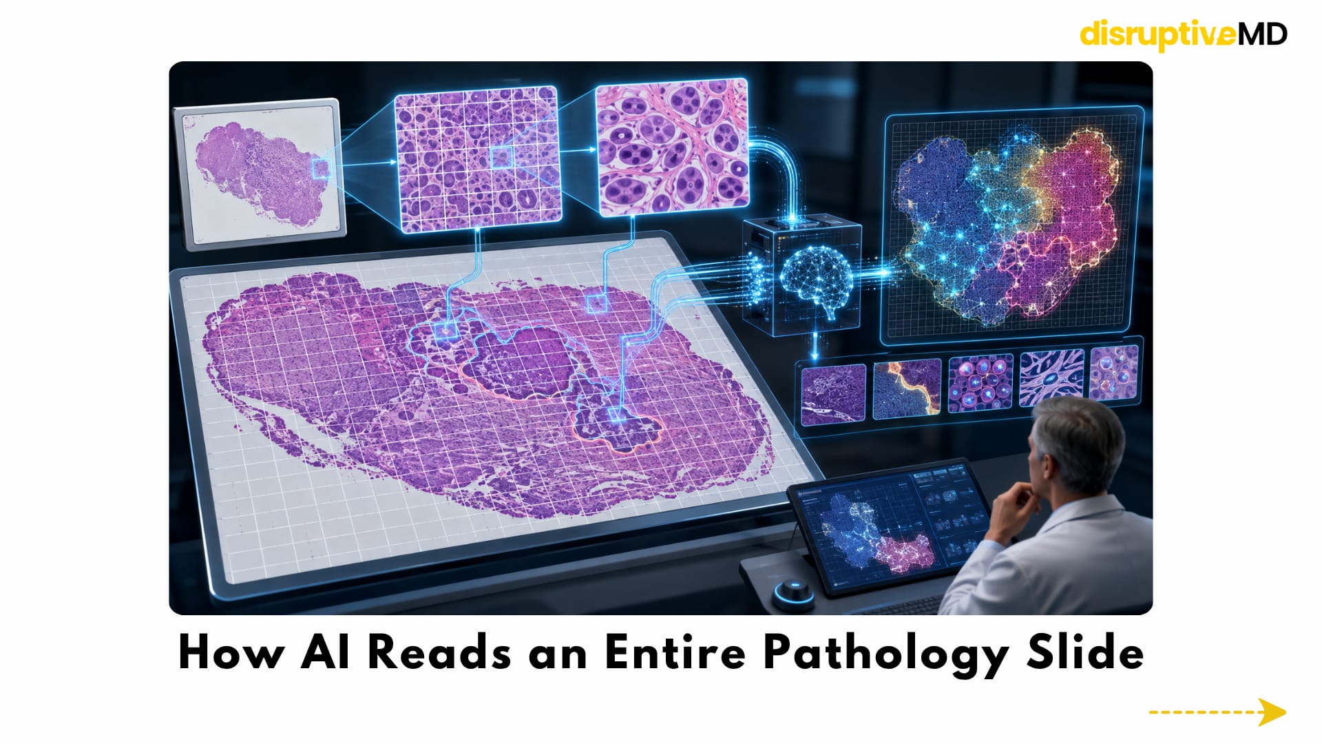
A whole-slide image is far larger than an ordinary photograph. It can contain billions of pixels and multiple levels of magnification.
An AI system usually cannot process the entire image as a single picture. Instead, the slide is divided into thousands of smaller regions called tiles or patches.
The model analyses these regions for features such as:
- abnormal cell shapes
- disrupted tissue architecture
- tumour–stroma boundaries
- gland formation
- nuclear irregularity
- immune-cell infiltration
- necrosis
- mitotic activity
- patterns of invasion
The results are then combined to produce a slide-level output.
Some systems use weak supervision, meaning that they learn from a diagnosis attached to the whole slide without requiring a pathologist to manually outline every malignant cell. Large-scale studies have shown that this approach can detect clinically meaningful patterns across whole-slide images while dramatically reducing the need for pixel-by-pixel annotation.
The model may also generate a heatmap that marks the regions contributing most strongly to its prediction.
This can help the pathologist understand where the system is focusing. However, a heatmap is not a complete explanation. It shows where the model looked, but it may not fully explain which biological feature drove the decision.
Finding the Tiny Focus That Changes the Diagnosis
One of AI pathology’s most immediate applications is cancer detection.
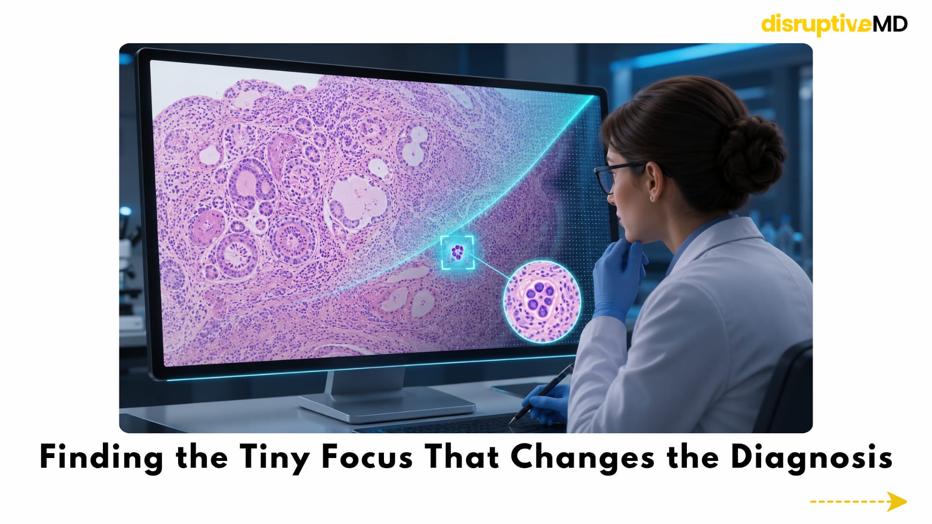
Some tissue samples contain large, obvious tumours. Others may contain only a few malignant glands, a microscopic deposit inside a lymph node or a tiny abnormal focus hidden among large areas of benign tissue.
That search is demanding because a pathologist may need to examine numerous slides containing millions of normal cells to find one clinically important region.
AI can act as a detection and triage system. It can scan the whole slide, identify suspicious areas and direct the pathologist’s attention towards regions that deserve closer inspection.
Research has demonstrated strong performance in selected applications such as:
- prostate biopsy assessment
- breast cancer lymph-node metastasis detection
- skin lesion classification
- gastrointestinal biopsy analysis
- tumour identification across several tissue types
In a large blinded prostate-biopsy study, an AI system demonstrated the ability to identify suspicious cancer regions within whole-slide images. Earlier comparative research also showed that high-performing algorithms could detect lymph-node metastases at a level comparable with pathologists under defined study conditions.
The most meaningful use, however, may not be AI working alone.
A more realistic model is pathologist plus AI.
In one workflow study, AI assistance reduced slide-reading time and improved metastasis detection for participating pathologists. The findings support the idea of AI as a second reader rather than an autonomous diagnostician.
Beyond Detection: Making Pathology Measurable
Pathology is not limited to deciding whether disease is present.
Many decisions depend on measurement.
A pathologist may need to determine:
- how many tumour cells express a biomarker
- how intense the staining appears
- how many cells are dividing
- how much of the tissue is tumour
- whether cancer approaches a surgical margin
- how many lymph nodes contain metastases
- how strongly immune cells have entered the tumour
These assessments are clinically important, but they can be affected by fatigue, borderline cases, tissue heterogeneity and differences between observers.
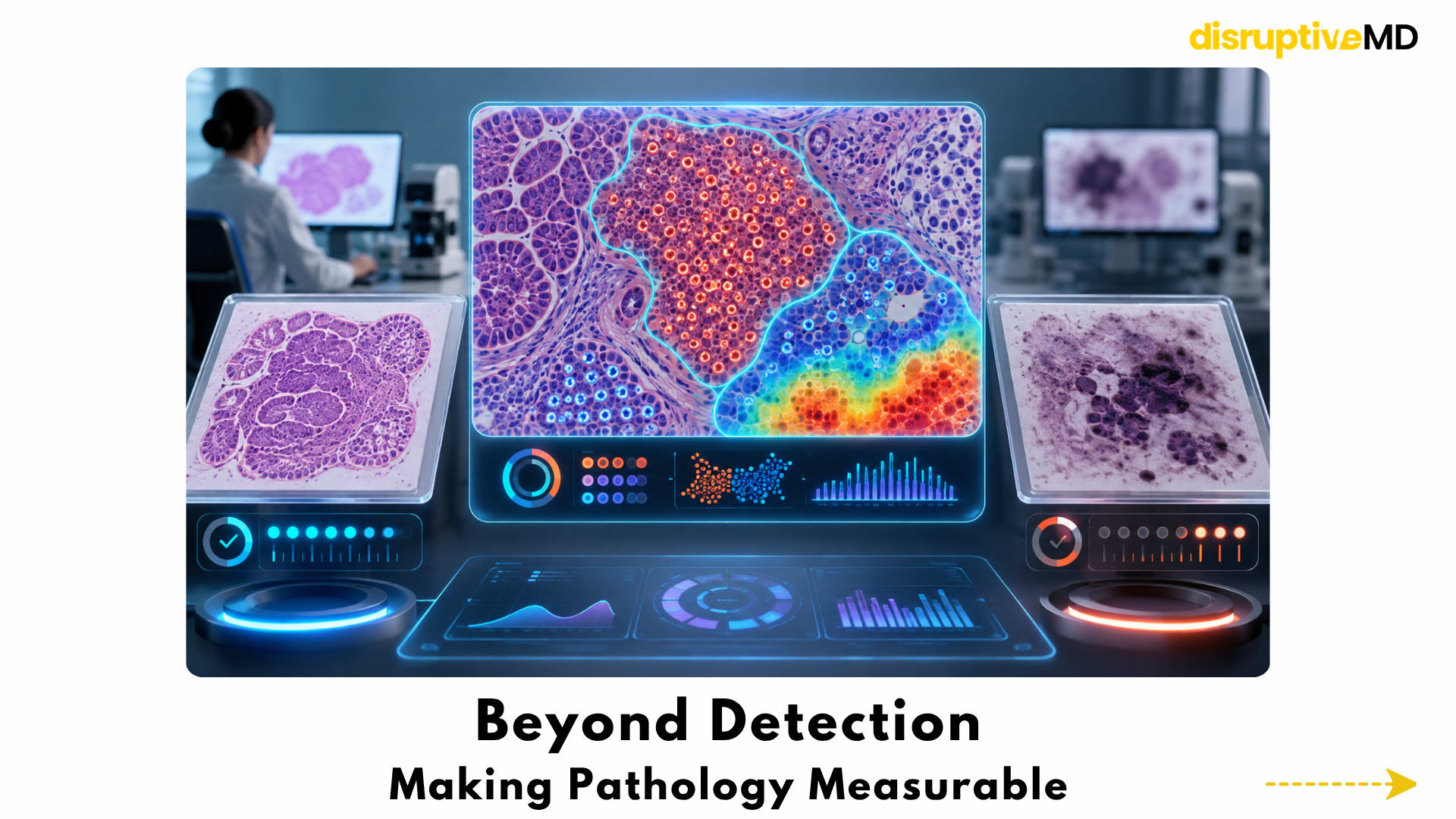
AI-assisted image analysis can count cells, segment tumour regions and measure staining across much larger tissue areas than manual sampling usually permits.
For example, algorithms are being studied for quantitative assessment of biomarkers such as Ki-67, hormone receptors, HER2 and PD-L1. In selected settings, digital measurement may improve consistency by separating tumour cells from surrounding tissue and calculating positivity across the relevant regions.
But an exact-looking number is not automatically an accurate number.
Results can still be affected by:
- tissue fixation
- staining quality
- antibody performance
- slide preparation
- scanner settings
- tumour heterogeneity
- incorrect tumour segmentation
AI can make pathology more quantitative, but the biological and technical quality of the specimen still determines the quality of the measurement.
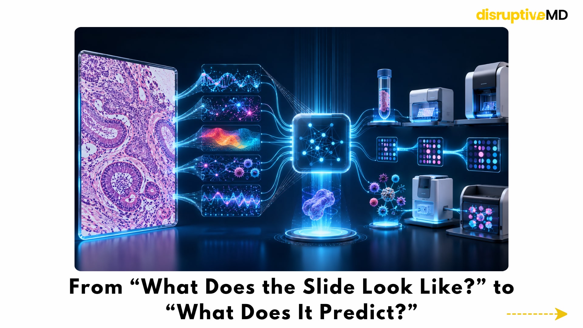
From “What Does the Slide Look Like?” to “What Does It Predict?”
The most futuristic direction in AI pathology is not simply finding cancer.
It is extracting hidden biological information from routine tissue images.
A standard stained slide contains visible patterns created by genes, proteins, metabolism, immune activity and interactions between tumour cells and their surrounding environment.
AI may be able to detect some of these patterns even when they are too subtle or complex to describe reliably by eye.
Research models have used routine pathology images to predict:
- selected genetic mutations
- microsatellite instability
- gene-expression patterns
- homologous recombination deficiency
- immune-cell composition
- tumour recurrence risk
- disease-specific survival
- possible treatment response
Early studies demonstrated that deep learning could classify major lung-cancer subtypes and predict selected mutations directly from tissue images. Other research showed that microsatellite instability and parts of a tumour’s gene-expression profile could be estimated from routinely stained slides.
AI has also been investigated for predicting survival across multiple cancer types and identifying histological patterns associated with aggressive disease.
This does not mean that a photograph of tissue can currently replace genomic sequencing or validated molecular testing.
Image-based predictions are indirect biological estimates. Their reliability can change across tumour types, laboratories and patient populations.
The near-term value may be as a screening or prioritisation layer. AI could identify cases with a higher probability of carrying a molecular feature, helping determine which samples require confirmatory testing first.
The slide may become a gateway to molecular information - not a substitute for it.
Pathology Foundation Models
Most early pathology algorithms were developed for one narrow task.
One model might detect prostate cancer. Another might count mitoses. Another might measure a biomarker.
A major new direction is the pathology foundation model.
A foundation model is trained on an extremely large and diverse collection of tissue images before being adapted to specific diagnostic tasks. Instead of learning only one cancer pattern, it learns broad visual representations of cells, tissue structures, inflammation, tumour architecture and disease.
Recent foundation models have been trained using:
- more than 100 million pathology image regions
- hundreds of thousands of whole-slide images
- over a billion individual tissue tiles
- multiple organs and cancer types
- paired pathology images and written reports
These models have shown the ability to adapt to cancer detection, tissue classification, biomarker prediction, rare-disease retrieval and prognosis-related tasks.
The newest multimodal models are learning not only from images but also from pathology reports and other clinical information. One large model reported in 2025 was trained on more than 335,000 whole-slide images using both visual learning and image–text alignment.
This creates the possibility of a general pathology intelligence system that can be adapted to many tasks without being rebuilt from the beginning each time.
But foundation models are not universal diagnostic engines.
Independent benchmarks have found that model performance varies substantially by task, dataset and clinical context. A model that performs strongly in cancer classification may not be the strongest option for biomarker prediction, prognosis or cell-level analysis.
Bigger does not automatically mean safer, more interpretable or more clinically useful.
Multimodal Pathology: Building a Complete Disease Picture
A tissue slide contains only one view of the disease.
The future of precision pathology may combine:
- whole-slide pathology images
- genomic and molecular information
- radiology scans
- blood biomarkers
- clinical history
- surgical findings
- previous treatments
- treatment response
- survival and recurrence data
A multimodal AI system could connect what the tumour looks like under the microscope with how it appears on imaging, which molecular pathways are active and how the disease has behaved over time.
For example, a radiology scan may show the location and spread of a tumour, while pathology reveals its cellular identity. Genomic information may reveal the molecular drivers. Blood markers may show how the disease is changing.
AI could help connect these separate layers into a patient-specific disease model.
This remains largely a research frontier. Combining different data types introduces missing information, incompatible formats, privacy concerns, timing differences and the danger that one unreliable input may distort the entire prediction.
The future may be multimodal, but every component will still require independent validation.
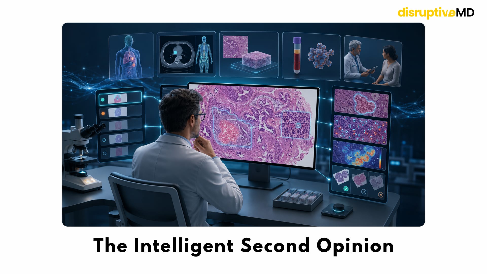
The Intelligent Second Opinion
The most realistic role for AI pathology is not an autonomous machine issuing a final diagnosis.
It is a tireless second reader.
AI could help with:
- prioritising urgent or suspicious slides
- highlighting possible tumour regions
- detecting small metastatic deposits
- counting tumour and immune cells
- measuring biomarkers
- identifying possible grading regions
- checking slide quality
- reviewing surgical margins
- flagging disagreement between images and reports
- supporting structured report preparation
This could allow the pathologist to spend less time on repetitive visual searches and more time on complex interpretation, clinical correlation and difficult cases.
The final diagnosis must remain a medical judgement.
A pathology report does not depend only on what appears in one image. It may require knowledge of the specimen, previous biopsies, imaging, surgery, laboratory findings and the clinical question being asked.
AI can analyse the slide.
The pathologist understands the case.
Why Heatmaps Are Not Enough
Clinical AI must be more than accurate on average.
It must also be understandable, reliable and capable of revealing uncertainty.
Heatmaps can show where a model found suspicious tissue, but they do not always explain the biological reasoning behind the prediction. A model may focus on a genuine tumour pattern, or on an irrelevant visual shortcut associated with a scanner, stain, tissue-processing method or laboratory.
This creates a dangerous possibility: an algorithm may appear highly accurate during development but fail when used on slides prepared differently.
Modern research has confirmed that even large pathology foundation models can encode technical differences between laboratories and scanners. Changes in tissue preparation, staining and slide acquisition can influence their internal representations and reduce performance outside the original environment.
Reliable clinical systems therefore need:
- external validation
- local laboratory testing
- monitoring after deployment
- clear confidence and failure indicators
- protection against automation bias
- human review of unexpected results
- procedures for scanner and stain changes
Explainability is not simply showing a colourful heatmap.
It is demonstrating that the prediction is based on real disease biology rather than an accidental feature of how the slide was produced.
The Risk of Bias
Pathology slides vary enormously.
Differences may arise from:
- scanner hardware
- staining protocols
- tissue thickness
- fixation time
- image compression
- laboratory workflow
- tumour subtype
- patient population
- disease prevalence
- the quality of manual annotations
An algorithm trained mostly on common tumours may perform poorly on rare variants. A model developed using one population may not generalise equally well to another. A system trained on carefully prepared research slides may struggle with folded tissue, blurred regions, air bubbles or incomplete scans encountered in routine practice.
Bias may also enter through the labels used to train the model.
If experts disagree about tumour grade, invasion or biomarker thresholds, the algorithm may learn that disagreement rather than eliminate it.
AI does not automatically remove subjectivity.
It can reproduce subjectivity at scale unless the training data, reference standards and validation process are carefully designed.
Fact Base
Strongest fact base
The strongest support exists for whole-slide digitisation and selected, narrowly defined AI tasks.
Digital pathology can support primary diagnostic review when the full workflow is properly validated. Task-specific algorithms have demonstrated strong performance in areas such as cancer detection, lymph-node screening and prostate-biopsy analysis. Studies also suggest that AI assistance can improve efficiency and sensitivity for pathologists in certain controlled workflows.
Developing fact base
Biomarker quantification, tumour grading, cell counting and workflow triage have meaningful clinical potential, but performance depends heavily on the disease, stain, scanner, laboratory and intended use.
AI may improve reproducibility, but it does not remove the need for specimen quality control or expert interpretation.
Emerging fact base
Predicting mutations, gene expression, prognosis and treatment response from routine slides is scientifically credible and supported by growing retrospective research.
However, many models still need prospective testing, external validation and proof that their predictions improve real treatment decisions rather than simply producing an additional risk score.
Research frontier
Foundation models, multimodal pathology, automated report generation and broadly generalisable AI systems represent the cutting edge.
They are advancing rapidly, but no single model can currently diagnose every disease, perform every pathology task or work equally well across all laboratories and patient populations. Current robustness studies continue to show vulnerability to technical variation.
What Is Real Today
What is real today:
- Glass slides can be converted into diagnostic-quality whole-slide images.
- Digital slides can support remote review and specialist collaboration.
- AI can assist with selected cancer-detection and screening tasks.
- Algorithms can highlight suspicious tissue using heatmaps.
- AI can count cells and support biomarker quantification.
- Selected systems are being integrated into regulated clinical workflows.
- Foundation models can be adapted to multiple research and diagnostic tasks.
- AI-assisted pathology can reduce repetitive work in carefully validated settings.
What is not fully real today: • AI replacing pathologists. • One universal model diagnosing every disease. • Fully automated tumour grading without expert review. • Molecular testing being routinely replaced by tissue-image prediction. • Autonomous treatment selection from a pathology slide. • Perfect performance across every scanner, stain, laboratory and population. • Fully automated pathology reports without human validation. • Heatmaps providing a complete explanation for every prediction.
Future Outlook The next stage of pathology will probably not look like a machine replacing a microscope.
It will look like a connected diagnostic workspace.
The pathologist may open a digital case and immediately see:
- areas prioritised for review
- possible tumour regions
- automated cell and biomarker measurements
- comparisons with previous specimens
- suspected molecular features
- quality-control warnings
- uncertainty estimates
- links between pathology, imaging and genomic information
- a structured draft requiring expert verification
The pathologist will decide which findings are meaningful, which are artefacts and which require additional testing.
As models improve, pathology may move beyond describing visible morphology towards measuring the tumour as a dynamic biological system.
The important transition is not from human intelligence to machine intelligence.
It is from isolated human inspection to human expertise supported by computational scale.
Key Takeaway
AI pathology is not about removing the human expert from diagnosis.
It is about giving the pathologist a second layer of intelligence—one that can scan, measure, compare, flag and predict across enormous digital slides.
The technology is already useful for selected tasks, but the most futuristic claims—universal diagnosis, molecular prediction and autonomous reporting—still require stronger validation.
The future pathologist may spend less time searching every part of the slide and more time interpreting what the tissue means for the patient.
The microscope is not disappearing. It is becoming intelligent.
Educational Disclaimer
This article is intended for medical and scientific education. It does not provide medical advice, recommend a diagnostic system or replace interpretation by a qualified pathology professional. AI performance varies according to the clinical task, tissue type, specimen quality, scanner, laboratory workflow and patient population.
References
- Mukhopadhyay S, et al. Whole Slide Imaging Versus Microscopy for Primary Diagnosis in Surgical Pathology: A Multicenter Blinded Randomized Noninferiority Study of 1992 Cases. American Journal of Surgical Pathology. 2018;42(1):39–52. DOI: 10.1097/PAS.0000000000000948.
- Ehteshami Bejnordi B, et al. Diagnostic Assessment of Deep Learning Algorithms for Detection of Lymph Node Metastases in Women With Breast Cancer. JAMA. 2017;318(22):2199–2210. DOI: 10.1001/jama.2017.14585.
- Campanella G, et al. Clinical-grade computational pathology using weakly supervised deep learning on whole slide images. Nature Medicine. 2019;25:1301–1309. DOI: 10.1038/s41591-019-0508-1.
- Pantanowitz L, et al. An artificial intelligence algorithm for prostate cancer diagnosis in whole slide images of core needle biopsies: a blinded clinical validation and deployment study. The Lancet Digital Health. 2020;2:e407–e416. DOI: 10.1016/S2589-7500(20)30159-X.
- Retamero JA, et al. Artificial Intelligence Helps Pathologists Increase Diagnostic Accuracy and Efficiency in the Detection of Breast Cancer Lymph Node Metastases. American Journal of Surgical Pathology. 2024;48(7):846–854. DOI: 10.1097/PAS.0000000000002248.
- Matsumoto H, et al. Ki-67 evaluation using deep-learning model-assisted digital image analysis in breast cancer. Histopathology. 2025;86:460–471. DOI: 10.1111/his.15356.
- Coudray N, et al. Classification and mutation prediction from non-small cell lung cancer histopathology images using deep learning. Nature Medicine. 2018;24:1559–1567. DOI: 10.1038/s41591-018-0177-5.
- Kather JN, et al. Deep learning can predict microsatellite instability directly from histology in gastrointestinal cancer. Nature Medicine. 2019;25:1054–1056. DOI: 10.1038/s41591-019-0462-y.
- Schmauch B, et al. A deep learning model to predict RNA-Seq expression of tumours from whole slide images. Nature Communications. 2020;11:3877. DOI: 10.1038/s41467-020-17678-4.
- Wulczyn E, et al. Deep learning-based survival prediction for multiple cancer types using histopathology images. PLOS ONE. 2020;15:e0233678. DOI: 10.1371/journal.pone.0233678.
- Chen RJ, et al. Towards a general-purpose foundation model for computational pathology. Nature Medicine. 2024;30:850–862. DOI: 10.1038/s41591-024-02857-3.
- Xu H, et al. A whole-slide foundation model for digital pathology from real-world data. Nature. 2024;630:181–188. DOI: 10.1038/s41586-024-07441-w.
- Vorontsov E, et al. A foundation model for clinical-grade computational pathology and rare cancers detection. Nature Medicine. 2024;30:2924–2935. DOI: 10.1038/s41591-024-03141-0.
- Campanella G, et al. A clinical benchmark of public self-supervised pathology foundation models. Nature Communications. 2025;16:3640. DOI: 10.1038/s41467-025-58796-1.
- Ding T, et al. A multimodal whole-slide foundation model for pathology. Nature Medicine. 2025;31:3749–3761. DOI: 10.1038/s41591-025-03982-3.
- Kömen J, et al. Towards robust foundation models for digital pathology. Nature Communications. 2026. DOI: 10.1038/s41467-026-73923-2.